Copyright
©2010 Baishideng Publishing Group Co.
World J Clin Oncol. Nov 10, 2010; 1(1): 3-11
Published online Nov 10, 2010. doi: 10.5306/wjco.v1.i1.3
Published online Nov 10, 2010. doi: 10.5306/wjco.v1.i1.3
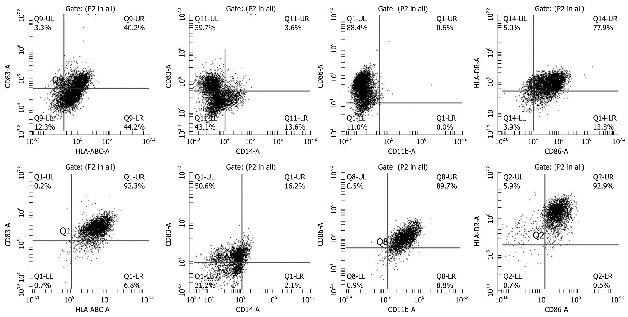
Figure 1 Phenotypic analysis of immature (upper panel) and mature dendritic cells (lower panel).
CD14+ cells collected from cord blood were incubated in immature dendritic cell (DC) media (RPMI-1640 plus 10% FBS) containing granulocyte colony stimulating factor (G-SCF) and IL-4 for 8 d and then cells were incubated with tumor necrosis factor (TNF)-α in addition to G-CSF and IL-4. Cells show typical monocytic DC markers (low CD14, high HLA-ABC, HLA-DR, CD86, CD83 and CD11b). Note the increase in the population of CD83 positive cells, a marker of mature DCs, after addition of TNF-α.
- Citation: Arbab AS. Cytotoxic T-cells as imaging probes for detecting glioma. World J Clin Oncol 2010; 1(1): 3-11
- URL: https://www.wjgnet.com/2218-4333/full/v1/i1/3.htm
- DOI: https://dx.doi.org/10.5306/wjco.v1.i1.3


