Copyright
©2014 Baishideng Publishing Group Inc.
World J Clin Oncol. Dec 10, 2014; 5(5): 1078-1087
Published online Dec 10, 2014. doi: 10.5306/wjco.v5.i5.1078
Published online Dec 10, 2014. doi: 10.5306/wjco.v5.i5.1078
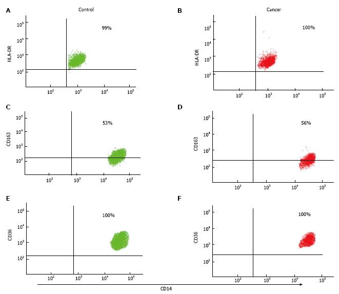
Figure 1 HLA-DR, CD163 and CD36 surface expression on (CD14+/CD16-) blood classical monocytes in patients with primary non-small cell lung cancer compared to controls.
A-F: Representative flow cytometry dot plots from PBMC stained against CD14, CD45 and CD16 and then co-stained with HLA-DR, CD163 and CD36 on patients with non-small cell lung cancer (red colour) and non-cancer controls (green colour).
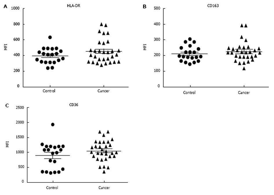
Figure 2 HLA-DR, CD163 and CD36 expression on (CD14+/CD16-) blood classical monocytes in patients with primary non-small cell lung cancer compared to controls.
Summary graphs show the mean values of MFI ± SEM of (A) HLA-DR, (B) CD163 and (C) CD36 markers from patients with non-small cell lung cancer (NSCLC) vs non-cancer controls.
Figure 3 CD11b, CD11c, CD71 and CD44 expression on (CD14+/CD16-) blood classical monocytes in patients with primary non-small cell lung cancer compared to controls.
Graphs show the mean values of MFI ± SEM of (A) CD11b, (B) CD11c, (C) CD71 and (D) CD44 markers from patients with non-small cell lung cancer vs non-cancer controls.
Figure 4 Th1 cytokine secretion profiles in plasma of patients with non-small cell lung cancer compared to controls.
Whole plasma was analysed for (A) TNF-α, (B) TNF-β, (C) IFN-γ, (D) IL-2, (E) IL-12 (p70) and (F) IL-1β by cytometric bead array technique using flow cytometery. Data was analysed using the FCAP Array™ v3.0.1 Software (BD Biosciences) and results are expressed as mean (pg/mL) ± SEM. IL: Interleukin; TNF: Tumor necrosis factor; IFN: Interferon.
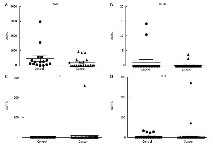
Figure 5 Th2 cytokine secretion profiles in plasma of patients with non-small cell lung cancer compared to controls.
Whole plasma was analysed for (A) IL-8, (B) IL-10, (C) IL-5 and (D) IL-4 by cytometric bead array technique using flow cytometery. Data was analysed using the FCAP Array™ v3.0.1 Software (BD Biosciences) and results are expressed as mean (pg/mL) ± SEM.
- Citation: Almatroodi SA, McDonald CF, Collins AL, Darby IA, Pouniotis DS. Blood classical monocytes phenotype is not altered in primary non-small cell lung cancer. World J Clin Oncol 2014; 5(5): 1078-1087
- URL: https://www.wjgnet.com/2218-4333/full/v5/i5/1078.htm
- DOI: https://dx.doi.org/10.5306/wjco.v5.i5.1078


