Copyright
©The Author(s) 2024.
World J Clin Oncol. Apr 24, 2024; 15(4): 566-575
Published online Apr 24, 2024. doi: 10.5306/wjco.v15.i4.566
Published online Apr 24, 2024. doi: 10.5306/wjco.v15.i4.566
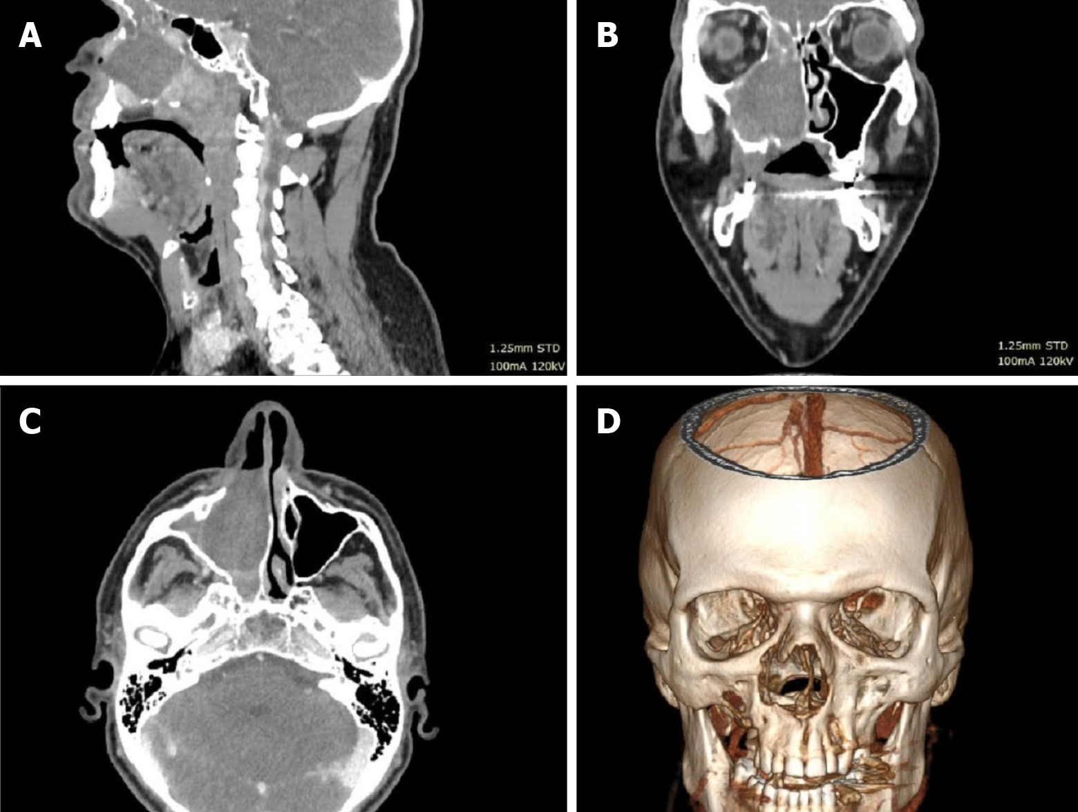
Figure 1 Case 1 computed tomography scan with contrast and 3D reconstruction.
Solid contrast-enhancing tumor filling the maxillary sinus and eroding the bony plate is shown. A: Sagittal section; B: Coronal section; C: Axial section; D: 3D reconstruction.
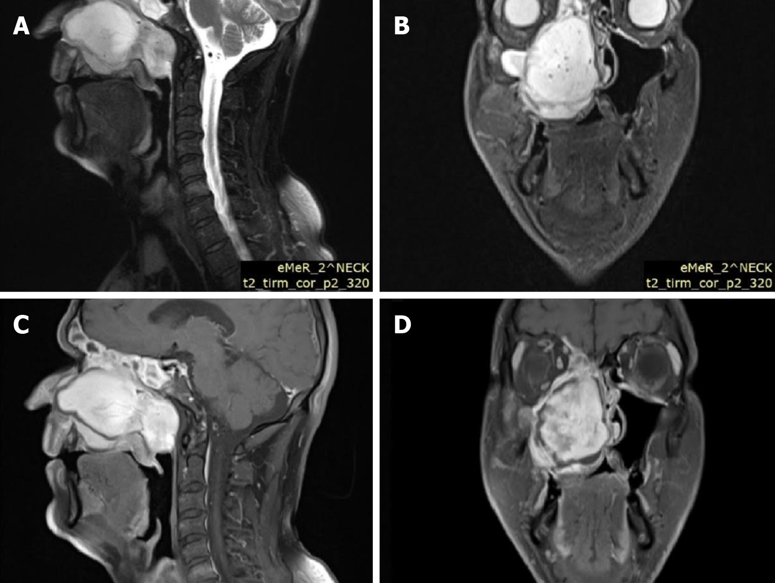
Figure 2 Case 1 T2 magnetic resonance imaging illustrating the extent of the tumor to the right maxilla sinus.
A and C: Sagittal sections; B and D: Coronal sections.
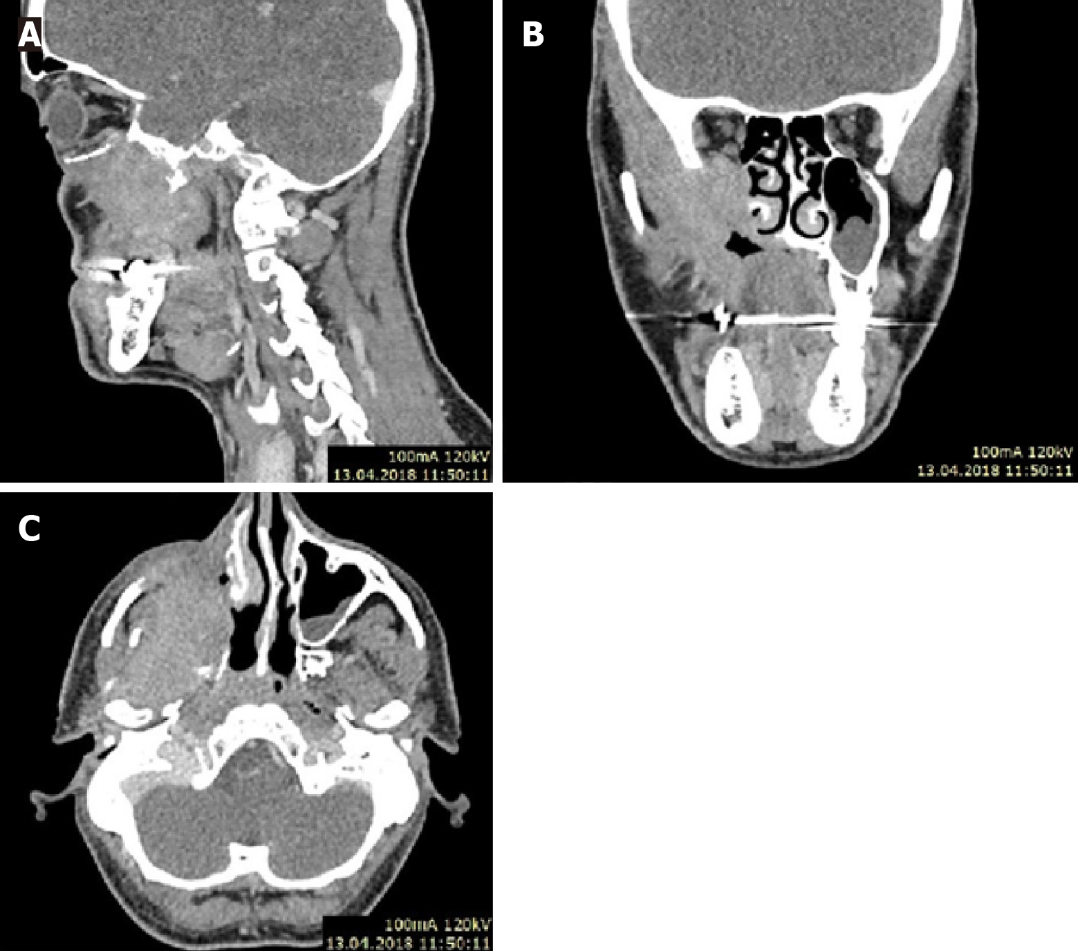
Figure 3 Case 2 computed tomography with contrast scan.
Solid contrast-enhancing tumor filling the maxillary sinus is shown. A: Sagittal section; B: Coronal section; C: Axial section.
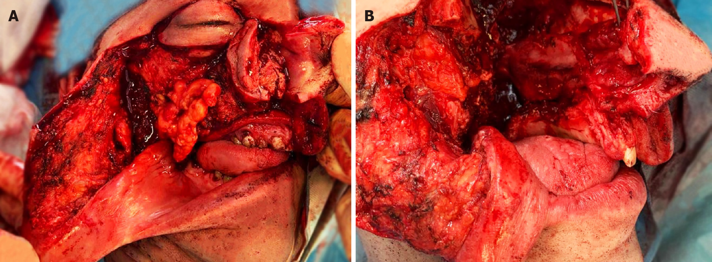
Figure 4 Case 1 intraoperative photo and post-resection lodge.
A: Intraoperative photo; B: Post-resection lodge.
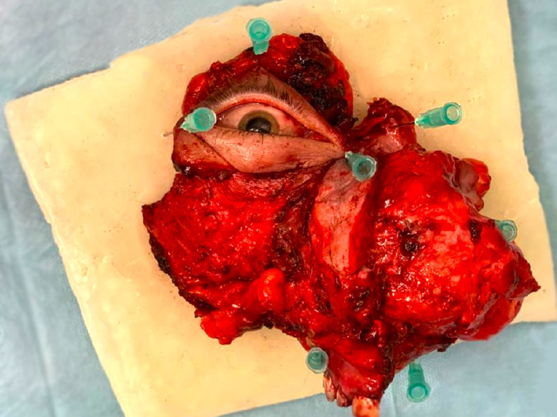
Figure 5
Case 1 fastened preparation in block.
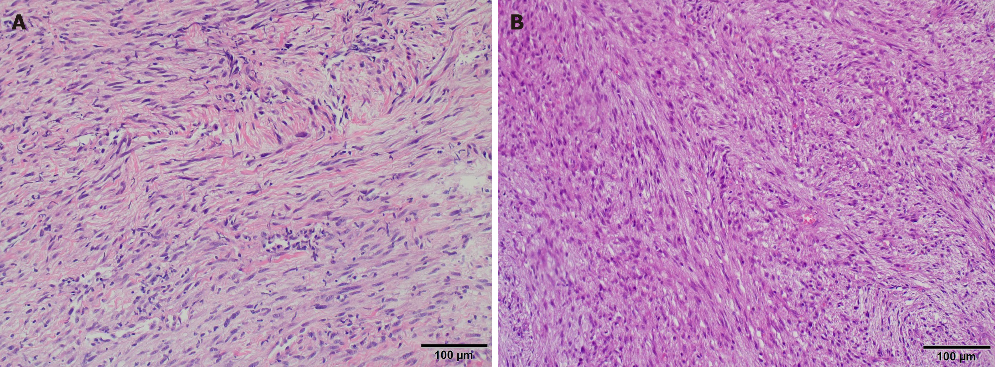
Figure 6 Hematoxylin and eosin staining.
A: Spindle cell infiltration, hypocellular with mild atypia, stromal collagen; hematoxylin and eosin (H&E) 20 ×; B: Hypercellular proliferation, fascicles of spindle cells; H&E 20 ×.
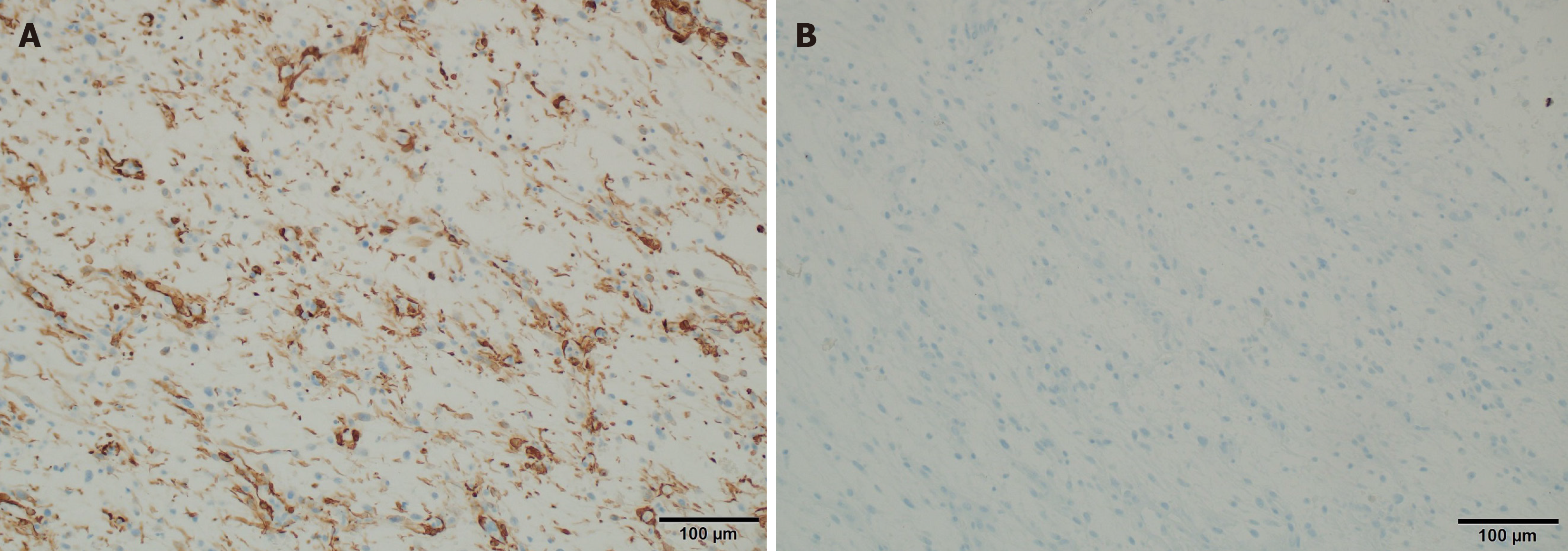
Figure 7 Focal expression of smooth muscle actin and no expression of anaplastic lymphoma kinase (magnification 20 ×).
A: Focal expression of smooth muscle actin (magnification 20 ×); B: No expression of anaplastic lymphoma kinase (magnification 20 ×).
- Citation: Mydlak A, Ścibik Ł, Durzynska M, Zwoliński J, Buchajska K, Lenartowicz O, Kucharz J. Low-grade myofibrosarcoma of the maxillary sinus: Two case reports. World J Clin Oncol 2024; 15(4): 566-575
- URL: https://www.wjgnet.com/2218-4333/full/v15/i4/566.htm
- DOI: https://dx.doi.org/10.5306/wjco.v15.i4.566


