Copyright
©The Author(s) 2022.
World J Clin Oncol. Oct 24, 2022; 13(10): 789-801
Published online Oct 24, 2022. doi: 10.5306/wjco.v13.i10.789
Published online Oct 24, 2022. doi: 10.5306/wjco.v13.i10.789
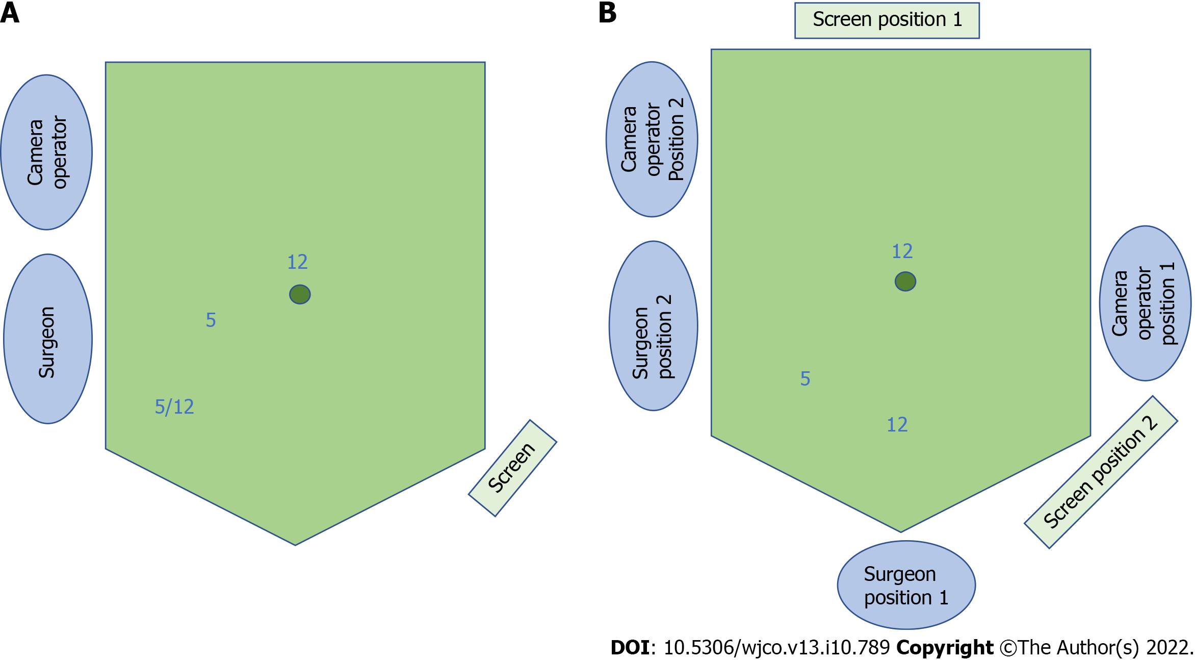
Figure 1 Schematic of 3-port natural orifice specimen extraction surgery port positioning and operative set-up.
A: For left-sided colorectal resection; the right iliac fossa port can be 5 or 12 mm depending on whether a linear stapler is used; B: For right-sided or subtotal/total colectomy, comprising position 1 for the initial phase of surgery and position 2 for the natural orifice specimen extraction procedure.
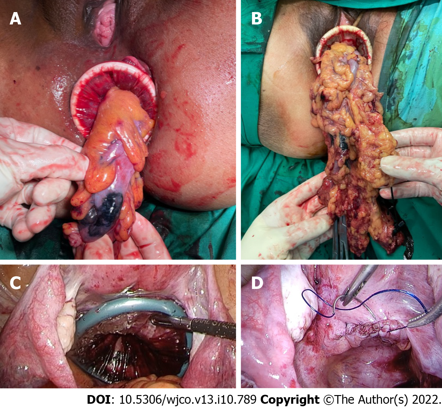
Figure 2 Operative images of the natural orifice specimen extraction procedure.
A: Transanal extraction; B: Transvaginal extraction; C: Intraperitoneal application of the dual-ring wound protector with the uterus hitched to the anterior abdominal wall; D: Closure of the posterior vaginotomy.
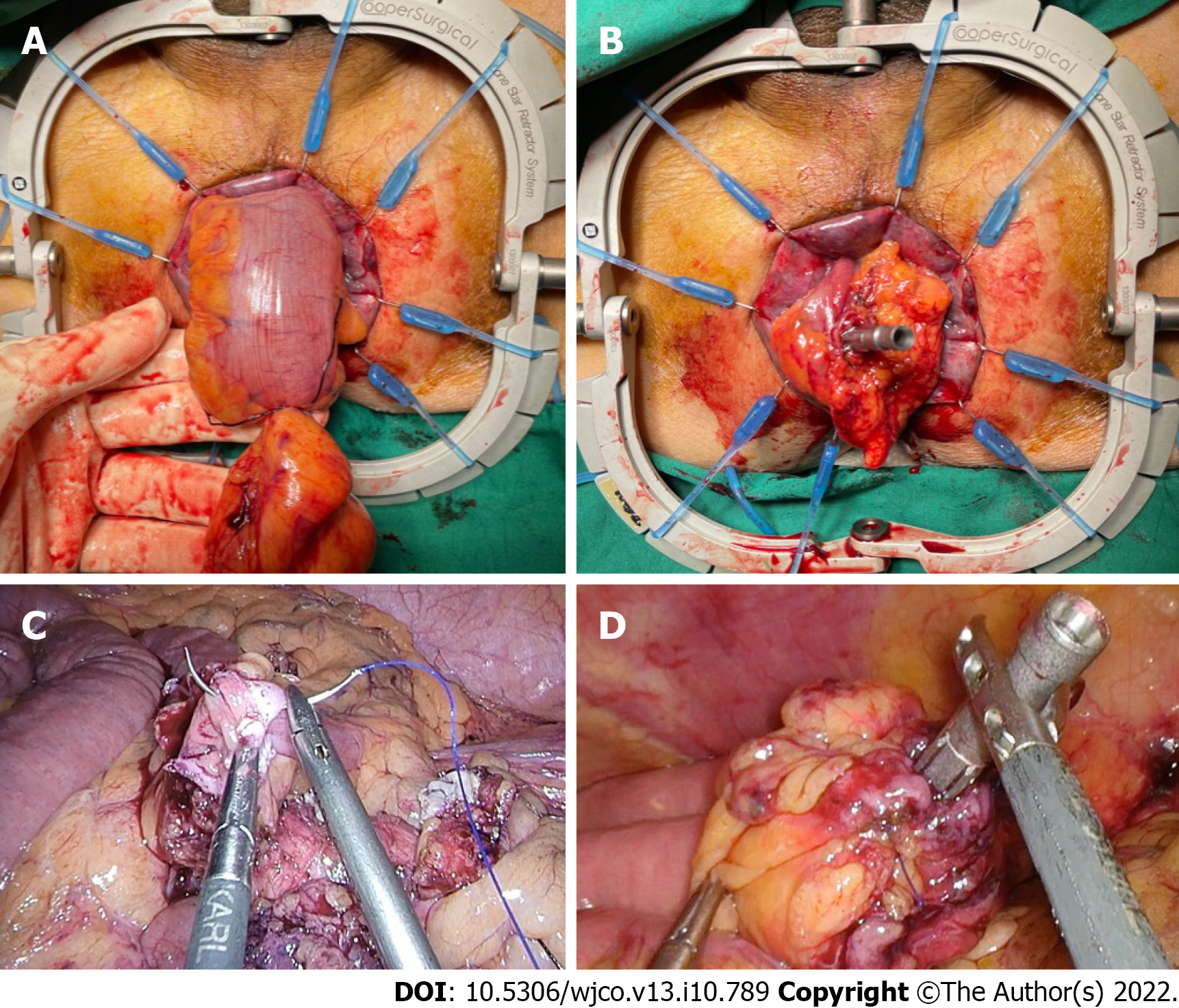
Figure 3 Methods of securing the circular stapler anvil to the proximal bowel.
A and B: Bowel pull-through and extracorporeal anvil application; C and D: Securing the anvil with an intracorporeal purse-string suture.
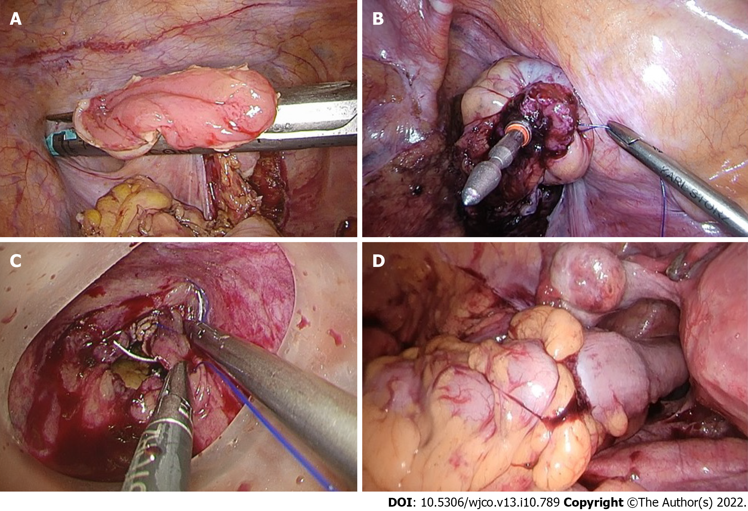
Figure 4 The hypothetical advantage of rectal purse-string closure is the creation of a double purse-string single-stapled anastomosis.
A and B: Methods of rectal stump closure linear-stapled closure, intracorporeal purse-string suture onto the fully extended spike of the circular stapler; C: Transanal purse-string suture with a transanal access device; D: A double purse-string single-stapled anastomosis.
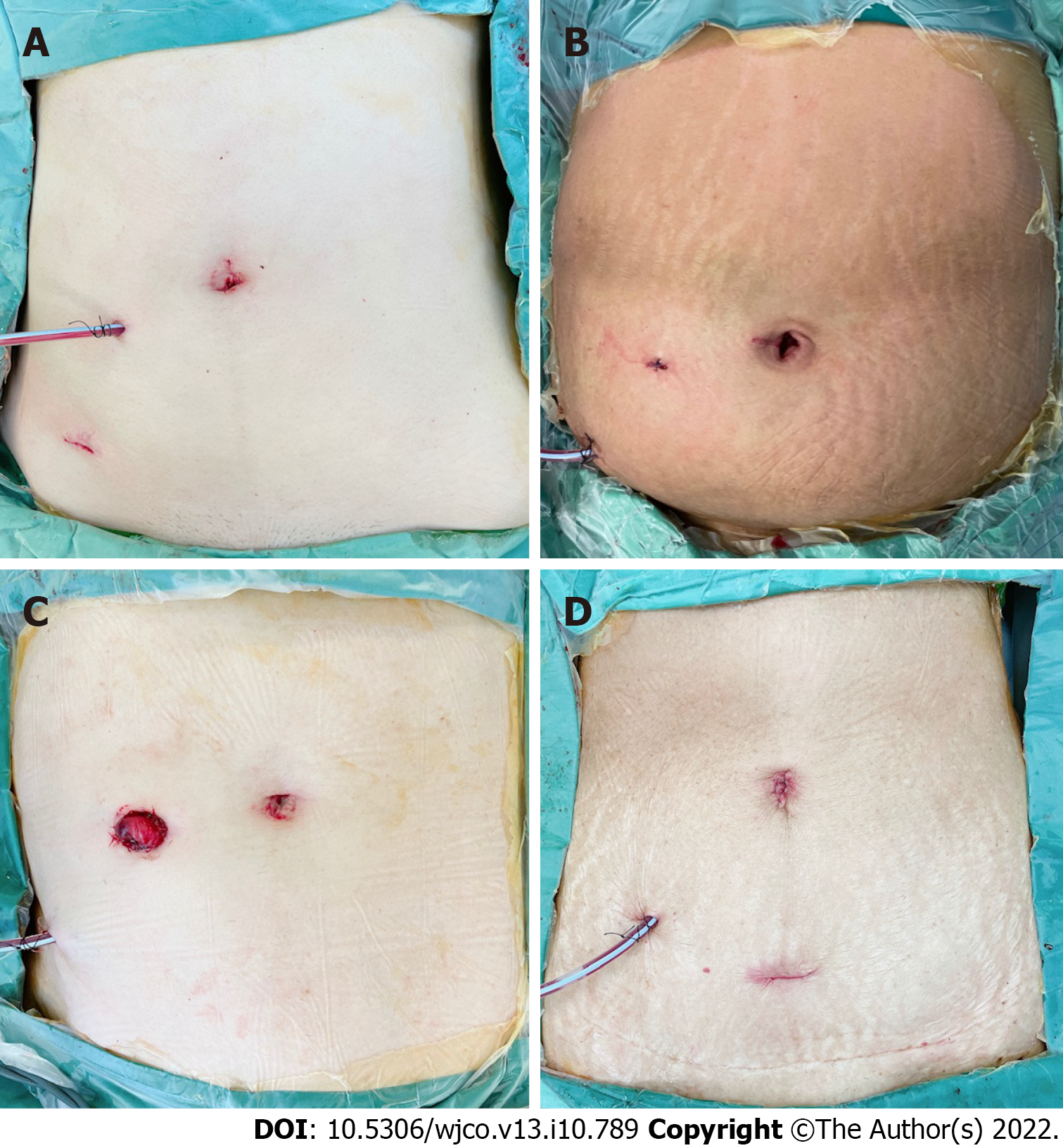
Figure 5 Postoperative abdominal incisions and appearance.
A: Anterior resection with transvaginal natural orifice specimen extraction (NOSE); B: Anterior resection with transanal NOSE; C: Low anterior resection and defunctioning ileostomy with transanal NOSE; D: D3 right hemicolectomy with transvaginal NOSE.
- Citation: Seow-En I, Chen LR, Li YX, Zhao Y, Chen JH, Abdullah HR, Tan EKW. Outcomes after natural orifice extraction vs conventional specimen extraction surgery for colorectal cancer: A propensity score-matched analysis. World J Clin Oncol 2022; 13(10): 789-801
- URL: https://www.wjgnet.com/2218-4333/full/v13/i10/789.htm
- DOI: https://dx.doi.org/10.5306/wjco.v13.i10.789


