Copyright
©The Author(s) 2015.
World J Biol Chem. Aug 26, 2015; 6(3): 83-94
Published online Aug 26, 2015. doi: 10.4331/wjbc.v6.i3.83
Published online Aug 26, 2015. doi: 10.4331/wjbc.v6.i3.83
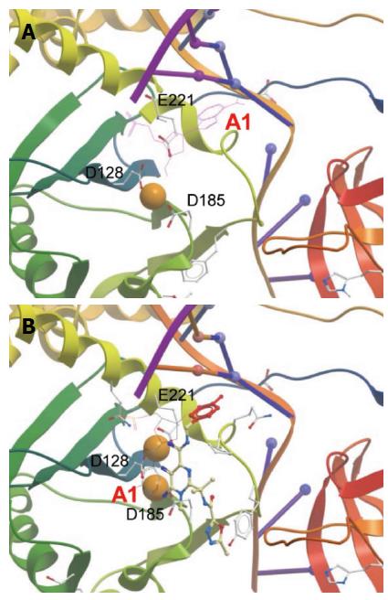
Figure 8 Active site within prototype foamy virus intasome in apo (A) and Raltegravir-bound state (B).
Orange spheres indicate Mg2+. Active sites residues are labeled. Only the terminal 3’ adenosine (marked A1) is displayed in its chemical form to show the displacement upon RAL binding. The diketo acid group of Raltegravir (RAL) interacts with the metal ions. Adenine is π-stacked against the RAL metal-chelating scaffold. The halobenzoyl group of RAL is marked red. The figures were constructed from PDB entries 3L2R and 3OYA, respectively, in Molsoft ICM-Browser.
- Citation: Grandgenett DP, Pandey KK, Bera S, Aihara H. Multifunctional facets of retrovirus integrase. World J Biol Chem 2015; 6(3): 83-94
- URL: https://www.wjgnet.com/1949-8454/full/v6/i3/83.htm
- DOI: https://dx.doi.org/10.4331/wjbc.v6.i3.83


