Copyright
©2013 Baishideng Publishing Group Co.
World J Biol Chem. Nov 26, 2013; 4(4): 131-140
Published online Nov 26, 2013. doi: 10.4331/wjbc.v4.i4.131
Published online Nov 26, 2013. doi: 10.4331/wjbc.v4.i4.131
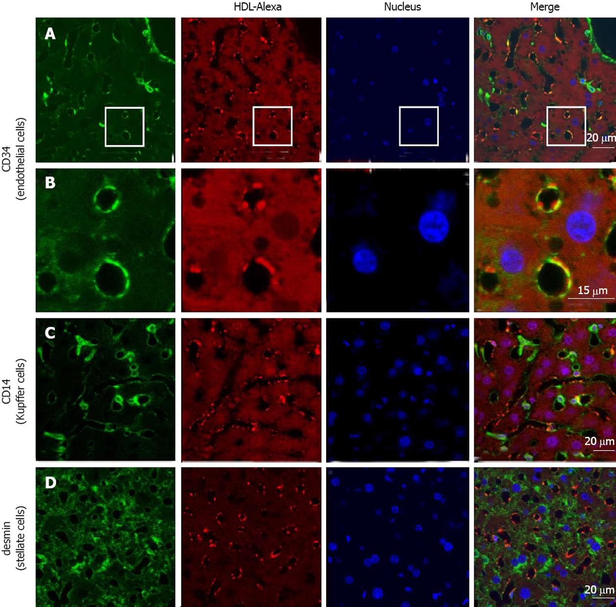
Figure 4 High density lipoprotein uptake in non-parenchymal liver cells.
Fluorescently labeled high density lipoprotein (HDL)-Alexa 568 was intravenously injected into C57BL/6 mice. After 60 min, tissues were fixed by transcardial perfusion. Liver sections were stained for cellular markers by immunofluorescence and analyzed by confocal microscopy. HDL is localized in endothelial cells [A and B (inset)] and to a limited amount in Kupffer cells (C), whereas stellate cells show no detectable HDL staining (D).
- Citation: Fruhwürth S, Pavelka M, Bittman R, Kovacs WJ, Walter KM, Röhrl C, Stangl H. High-density lipoprotein endocytosis in endothelial cells. World J Biol Chem 2013; 4(4): 131-140
- URL: https://www.wjgnet.com/1949-8454/full/v4/i4/131.htm
- DOI: https://dx.doi.org/10.4331/wjbc.v4.i4.131


