Copyright
©2012 Baishideng Publishing Group Co.
World J Biol Chem. May 26, 2012; 3(5): 98-109
Published online May 26, 2012. doi: 10.4331/wjbc.v3.i5.98
Published online May 26, 2012. doi: 10.4331/wjbc.v3.i5.98
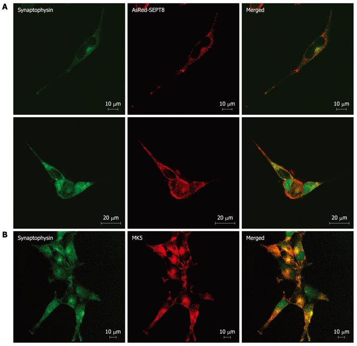
Figure 5 Mitogen-activated protein kinase-activated protein kinase 5 and SEPT8 colocalize with synaptophysin.
A: SK-N-DZ cells were transfected with expression vector encoding AsRed-SEPT8 and after 24 h, cells were fixed. Synaptophysin was visualized by staining with anti-synaptophysin fluorescein isothiocyanate (FITC) conjugate antibody (left panels), while AsRed-SEPT8 was visualized directly (red channel; middle panels). A merged image of red and green channels is shown in the right panel; B: SK-N-DZ cells were fixed and stained with anti-synaptophysin FITC conjugate antibody (left panel) and with anti-PRAK antibody followed by Alexa Fluor 568 anti-rabbit antibody (middle panel). Merged image of red and green channels is shown in the right panel.
-
Citation: Shiryaev A, Kostenko S, Dumitriu G, Moens U. Septin 8 is an interaction partner and
in vitro substrate of MK5. World J Biol Chem 2012; 3(5): 98-109 - URL: https://www.wjgnet.com/1949-8454/full/v3/i5/98.htm
- DOI: https://dx.doi.org/10.4331/wjbc.v3.i5.98


