Copyright
©2011 Baishideng Publishing Group Co.
World J Biol Chem. Oct 26, 2011; 2(10): 232-238
Published online Oct 26, 2011. doi: 10.4331/wjbc.v2.i10.232
Published online Oct 26, 2011. doi: 10.4331/wjbc.v2.i10.232
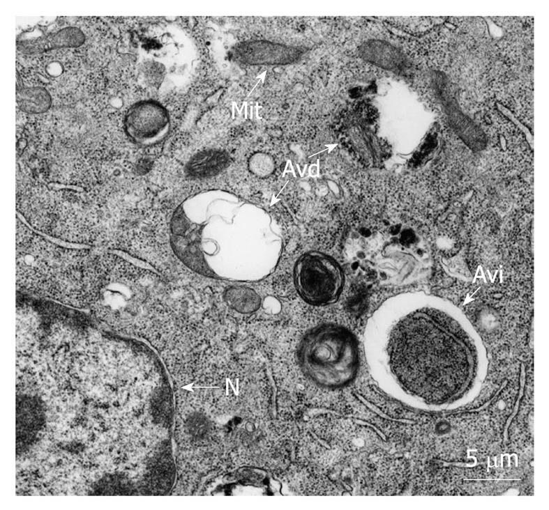
Figure 1 Morphology of autophagic vacuoles.
Typical autophagic vacuoles from 3T3 mouse fibroblasts incubated in a nutrient-poor medium containing cytoplasmic material at early (Avi) and late (Avd) degradation stages. Mit: Mitochondria; N: Nucleus.
- Citation: Esteve JM, Knecht E. Mechanisms of autophagy and apoptosis: Recent developments in breast cancer cells. World J Biol Chem 2011; 2(10): 232-238
- URL: https://www.wjgnet.com/1949-8454/full/v2/i10/232.htm
- DOI: https://dx.doi.org/10.4331/wjbc.v2.i10.232


