
















Copyright: ©Author(s) 2026.
World J Biol Chem. Sep 5, 2026; 17(3): 119936
Published online Sep 5, 2026. doi: 10.4331/wjbc.119936
Published online Sep 5, 2026. doi: 10.4331/wjbc.119936
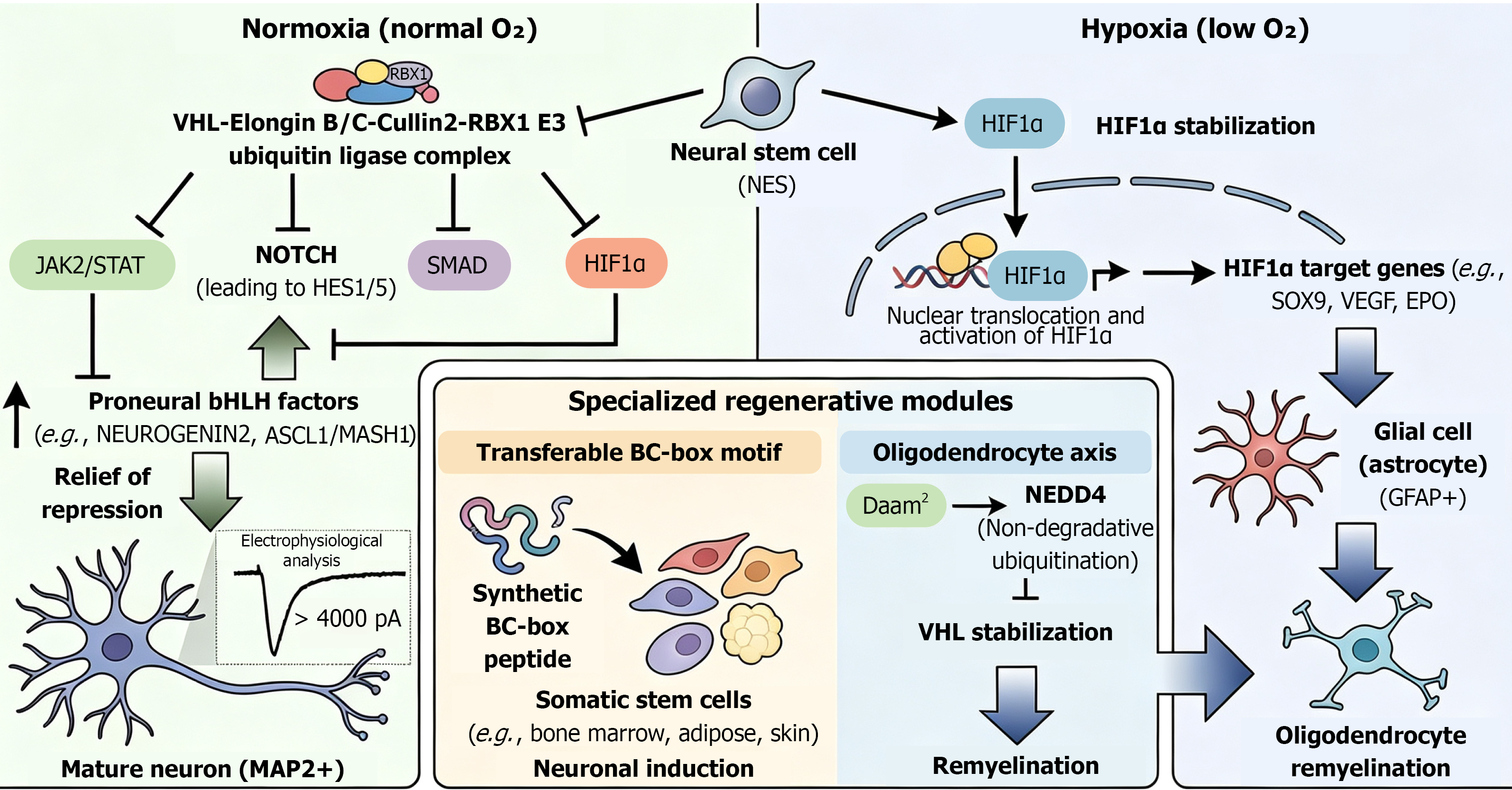
Figure 1 Regenerative potential and molecular mechanisms of the von Hippel-Lindau protein in neural and somatic stem cells.
Normoxia (Left): Under normal oxygen conditions, von Hippel-Lindau (VHL) functions as the substrate-recognition component of the VHL-Elongin B/C-Cullin2-RBX1 E3 ubiquitin ligase complex. This complex targets several key signaling molecules for proteasomal degradation, including JAK2/STAT, NOTCH (HES1/5), SMAD, and HIF-1α. The suppression of these pathways leads to the “relief of repression” for proneural bHLH factors (e.g., NEUROGENIN2, ASCL1/MASH1), thereby promoting the differentiation of neural stem cells (NSCs) into functional, mature neurons (MAP2+) characterized by robust electrophysiological activity (> 4000 pA). Hypoxia (right): Under low oxygen conditions, VHL-mediated degradation is inhibited, leading to HIF1α stabilization. Stabilized HIF-1α translocates to the nucleus and activates target genes such as SOX9, VEGF, and EPO. This pathway shifts the NSC differentiation bias toward the glial lineage, specifically promoting the formation of GFAP+ astrocytes. Specialized regenerative modules (bottom center): Transferable BC-Box motif: A synthetic BC-box peptide can mimic VHL function to induce neuronal differentiation in various somatic stem cells (e.g., bone marrow, adipose, and skin-derived cells), suggesting a modular application for regenerative medicine. Oligodendrocyte axis: The Daam2-NEDD4 axis regulates VHL via non-degradative ubiquitination. VHL stabilization through this pathway is critical for remyelination, facilitating the maturation of oligodendrocytes and the repair of myelin sheaths. VHL: Von Hippel-Lindau; NSCs: Neural stem cells.
- Citation: Kanno H, Nakahara K. Regenerative potential of von Hippel-Lindau tumor suppressor protein. World J Biol Chem 2026; 17(3): 119936
- URL: https://www.wjgnet.com/1949-8454/full/v17/i3/119936.htm
- DOI: https://dx.doi.org/10.4331/wjbc.119936