Copyright
©The Author(s) 2023.
World J Biol Chem. Oct 17, 2023; 14(5): 84-98
Published online Oct 17, 2023. doi: 10.4331/wjbc.v14.i5.84
Published online Oct 17, 2023. doi: 10.4331/wjbc.v14.i5.84
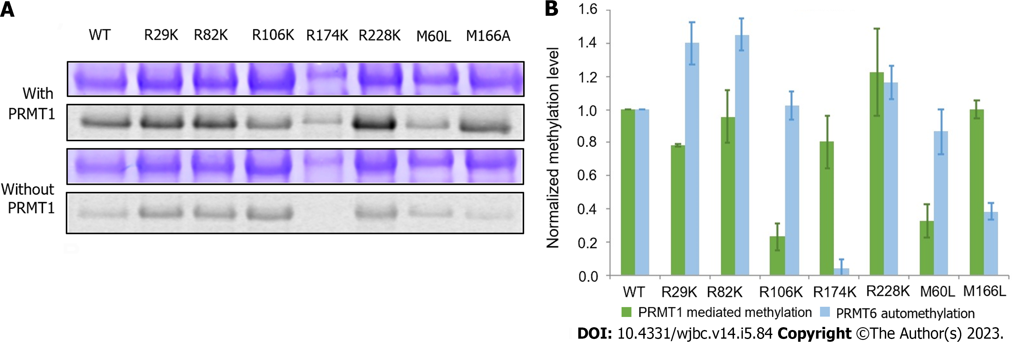
Figure 5 Comparison of protein arginine methyltransferase 1-mediated methylation and automethylation among different protein arginine methyltransferase 6 mutants.
A: Radioactive methyltransferase assay was performed to compare protein arginine methyltransferase (PRMT) 1-mediated methylation level and automethylation level of PRMT6 mutants. 5 M of each PRMT6 mutant was incubated with 30 M 14C-S-adenosyl methionine in the presence or absence of 1 M PRMT1 in the reaction buffer at 30 ℃ for 2 h. The products were separated on 12% sodium dodecyl sulfate polyacrylamide gel electrophoresis and visualized by Coomassie blue staining and phosphor image scanning. The methylation intensity of each mutant was quantified with Quantity One and normalized against PRMT6WT. The assay was repeated at least three times. Coomassie blue staining and phosphor image of methylated PRMT6 mutants in the presence or absence of PRMT1; B: Column graph comparing the methylation of PRMT6 m. PRMT: Protein arginine methyltransferase.
- Citation: Cao MT, Feng Y, Zheng YG. Protein arginine methyltransferase 6 is a novel substrate of protein arginine methyltransferase 1. World J Biol Chem 2023; 14(5): 84-98
- URL: https://www.wjgnet.com/1949-8454/full/v14/i5/84.htm
- DOI: https://dx.doi.org/10.4331/wjbc.v14.i5.84


