Copyright
©The Author(s) 2023.
World J Biol Chem. Oct 17, 2023; 14(5): 84-98
Published online Oct 17, 2023. doi: 10.4331/wjbc.v14.i5.84
Published online Oct 17, 2023. doi: 10.4331/wjbc.v14.i5.84
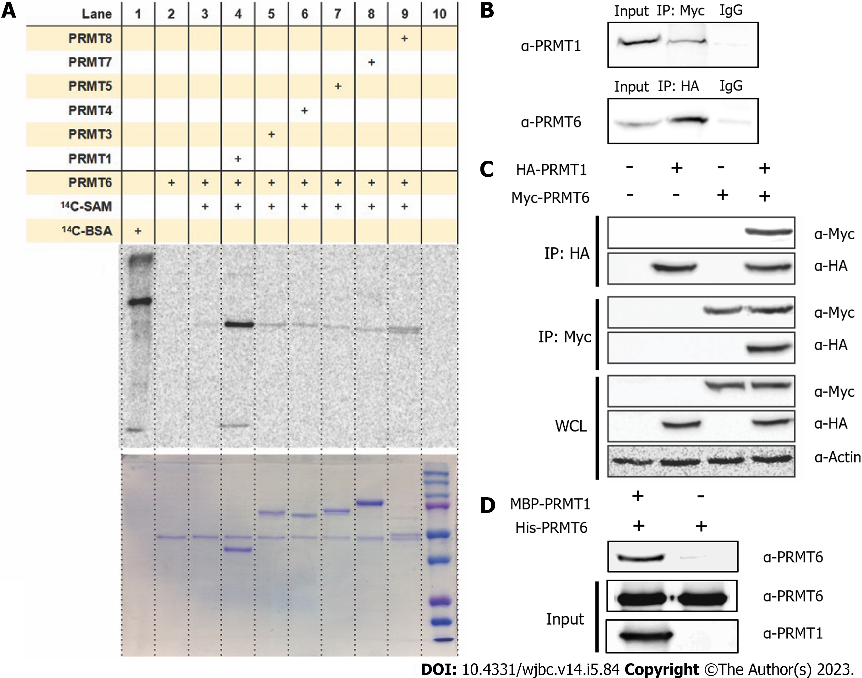
Figure 1 Protein arginine methyltransferase 1 interacts with protein arginine methyltransferase 6 in vitro and in vivo.
A: Gel electrophoresis-autoradiographic image analysis of protein arginine methyltransferase (PRMT) 6 methylation incubated with other PRMT family members. 0.5 µM of PRMT6 was incubated with 1 µM of PRMT1, PRMT3, coactivator associated arginine methyltransferase (PRMT4), PRMT5, PRMT7 and PRMT8. Reactions were held for 30 min at 30 ℃, the samples were resolved by sodium dodecyl sulfate-polyacrylamide gel electrophoresis (SDS-PAGE) with a 12% polyacrylamide gel; B: Co-immunoprecipitation assay was performed between HA-PRMT1 and endogenous PRMT6 or Myc-PRMT6 and endogenous PRMT1 in HEK293T cells. Co-transfected HEK293T cell lysate was immunoprecipitated with anti-HA antibody or anti-Myc antibody followed by protein A/G plus agarose beads, proteins on the beads were eluted and separated on 12% SDS-PAGE and were detected by anti-PRMT1 or anti-PRMT6 antibody; C: Immunoprecipitation assay was performed between HA-PRMT1 and Myc-PRMT6. Single transfected HEK293T Cell lysate was immunoprecipitated with anti-HA antibody or anti-Myc antibody followed by protein A/G plus agarose beads, proteins on the beads were eluted and separated on 12% SDS-PAGE and were detected by anti-Myc or anti-HA antibody; D: Western blot analysis of maltose-binding protein (MBP) pull down assay. PRMT6 binds to purified Amylose beads or MBP-PRMT1 was detected. PRMT1 was incubated with the amylose beads for 30 min, followed by washing by phosphate-buffered saline and incubated with PRMT6 for 4h. The resin was washed and the pulled-down proteins were eluted and separated by 12% SDS-PAGE and detected by anti-PRMT1 or anti-PRMT6. IgG: Immunoglobulin G; PRMT: Protein arginine methyltransferase; SAM: S-adenosyl methionine.
- Citation: Cao MT, Feng Y, Zheng YG. Protein arginine methyltransferase 6 is a novel substrate of protein arginine methyltransferase 1. World J Biol Chem 2023; 14(5): 84-98
- URL: https://www.wjgnet.com/1949-8454/full/v14/i5/84.htm
- DOI: https://dx.doi.org/10.4331/wjbc.v14.i5.84


