Copyright
©The Author(s) 2022.
World J Biol Chem. Mar 27, 2022; 13(2): 47-65
Published online Mar 27, 2022. doi: 10.4331/wjbc.v13.i2.47
Published online Mar 27, 2022. doi: 10.4331/wjbc.v13.i2.47
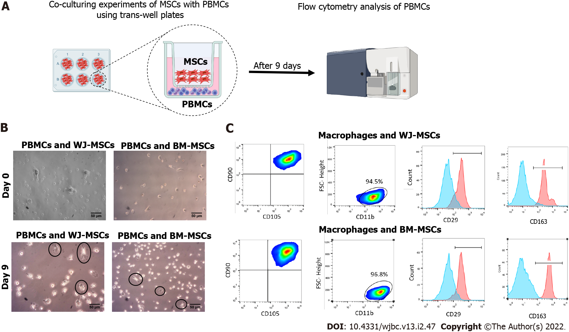
Figure 5 Co-culturing experiments of M1 macrophages with stimulated Wharton’s Jelly and bone marrow-mesenchymal stromal cells.
A: Schematic representation of the experimental workflow; B: Macrophage morphology was changed 9 d after co-culture either with Wharton’s Jelly or bone marrow-mesenchymal stromal cells. After 9 d, macrophages exhibited a more elongated shape and plastic adherence. Black arrows indicate the presence of elongated plastic adherent cells; C: Flow cytometry analysis showed the positive expression of CD45, CD14, and CD11b by the differentiated macrophages. After 9 d, the macrophages exhibited an increase in the CD29 and CD163 expression, compared to the cells at day 0. Statistically significant differences regarding the CD29 and CD163 were observed in M2 compared to M1 macrophages (P < 0.001). BM: Bone marrow; MSCs: Mesenchymal stromal cells; WJ: Wharton’s Jelly; PBMCs: Peripheral blood mononuclear cells.
- Citation: Mallis P, Chatzistamatiou T, Dimou Z, Sarri EF, Georgiou E, Salagianni M, Triantafyllia V, Andreakos E, Stavropoulos-Giokas C, Michalopoulos E. Mesenchymal stromal cell delivery as a potential therapeutic strategy against COVID-19: Promising evidence from in vitro results. World J Biol Chem 2022; 13(2): 47-65
- URL: https://www.wjgnet.com/1949-8454/full/v13/i2/47.htm
- DOI: https://dx.doi.org/10.4331/wjbc.v13.i2.47


