Copyright
©The Author(s) 2022.
World J Biol Chem. Mar 27, 2022; 13(2): 47-65
Published online Mar 27, 2022. doi: 10.4331/wjbc.v13.i2.47
Published online Mar 27, 2022. doi: 10.4331/wjbc.v13.i2.47
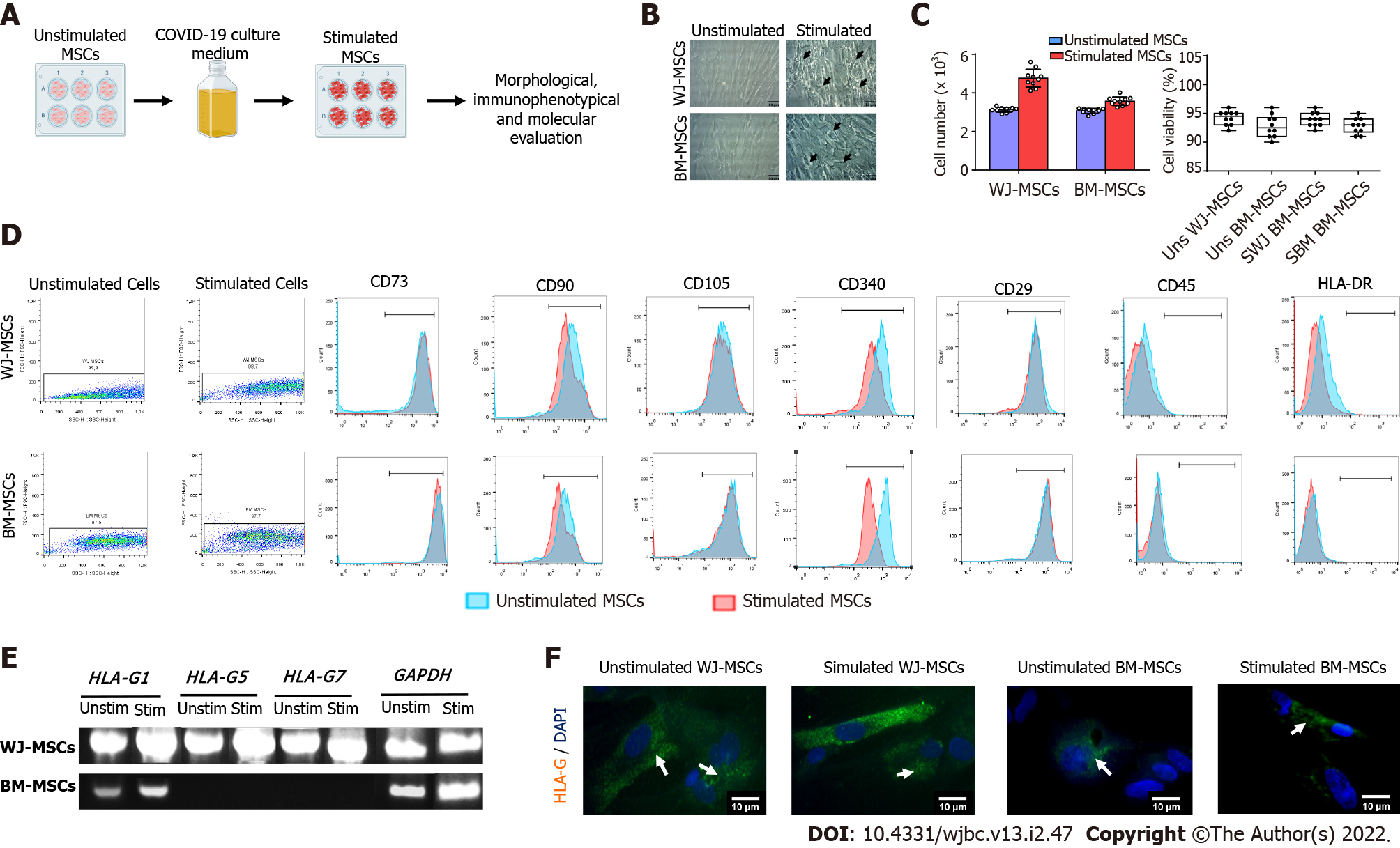
Figure 2 Comprehensive characterization of characteristics of stimulated Wharton’s Jelly and bone marrow mesenchymal stromal cells.
A: Experimental workflow; B: Morphological analysis of characteristics of unstimulated and stimulated Wharton’s Jelly (WJ) and bone marrow-mesenchymal stromal cells (BM-MSCs). Original magnification 20 ×, scale bars = 50 μm; C: Determination of cell proliferation and viability. Statistically significant differences were observed in cell proliferation between stimulated and unstimulated WJ-MSCs (P < 0.05) and stimulated and unstimulated BM-MSCs (P < 0.05). No statistically significant difference was observed in cell viability either in an unstimulated or stimulated state (P = 0.873); D: Immunophenotypic analysis of stimulated and unstimulated WJ and BM-MSCs. Over 95% of WJ and BM-MSCs in both states expressed CD73, CD90, CD105, CD29, and CD340, and less than 2% expressed CD34 and CD45; E: Determination of HLA-G isoforms (HLA-G1, G5, and G7) in unstimulated and stimulated MSCs from both sources; F: Indirect immunofluorescence against HLA-G1 in combination with DAPI stain was performed on unstimulated and stimulated WJ and BM-MSCs. Original magnification 63 ×, scale bars = 10 μm. BM: Bone marrow; MSCs: Mesenchymal stromal cells; WJ: Wharton’s Jelly.
- Citation: Mallis P, Chatzistamatiou T, Dimou Z, Sarri EF, Georgiou E, Salagianni M, Triantafyllia V, Andreakos E, Stavropoulos-Giokas C, Michalopoulos E. Mesenchymal stromal cell delivery as a potential therapeutic strategy against COVID-19: Promising evidence from in vitro results. World J Biol Chem 2022; 13(2): 47-65
- URL: https://www.wjgnet.com/1949-8454/full/v13/i2/47.htm
- DOI: https://dx.doi.org/10.4331/wjbc.v13.i2.47


