Copyright
©The Author(s) 2021.
World J Biol Chem. Jan 27, 2021; 12(1): 1-14
Published online Jan 27, 2021. doi: 10.4331/wjbc.v12.i1.1
Published online Jan 27, 2021. doi: 10.4331/wjbc.v12.i1.1
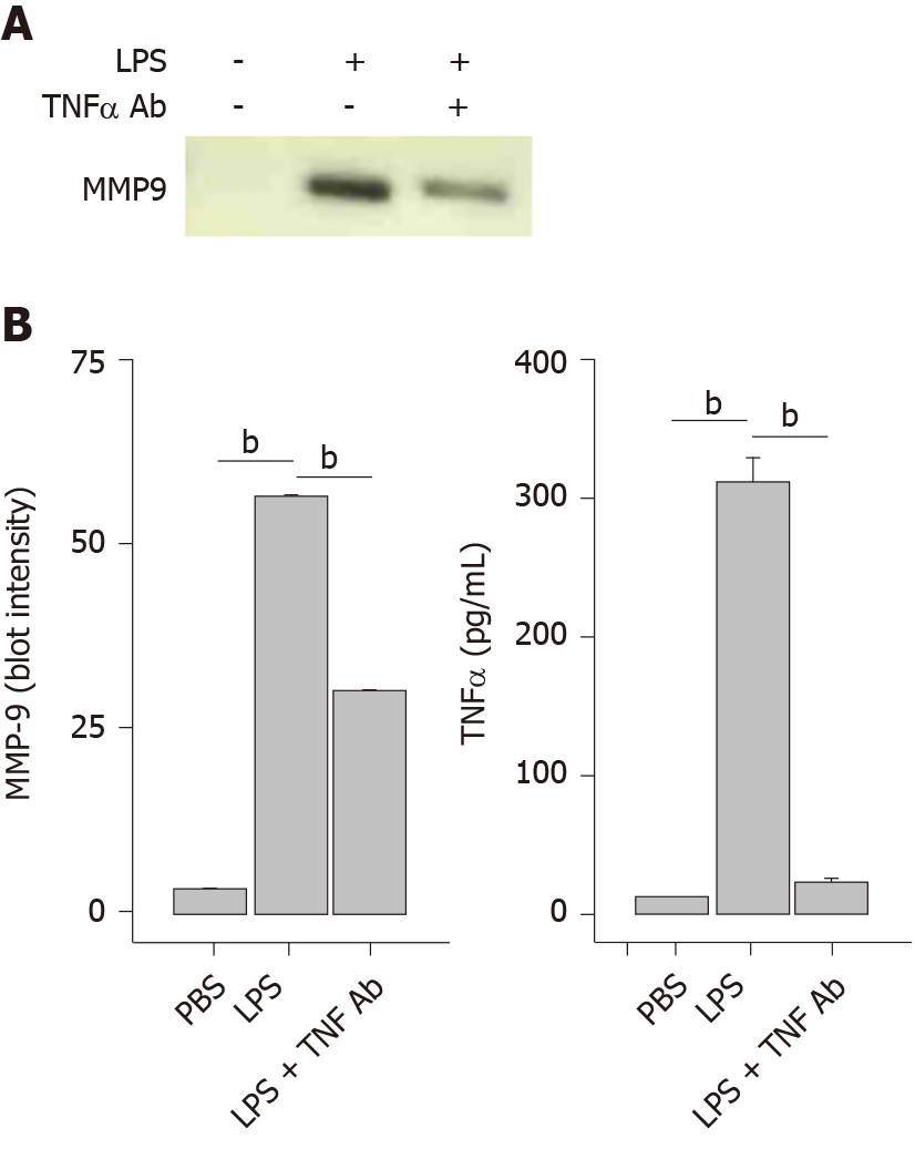
Figure 6 Antif- tumor necrosis factor α antibody blocks lipopolysaccharide-induced matrix-metalloproteinase-9 secretion.
THP-1 monocytes were treated with a tumor necrosis factor α (TNFα) antibody (1 μg/mL) followed by lipopolysaccharide (1 μg/mL) for 48 h. Cells were collected, centrifuged at 500 g for 10 min and supernatant assessed for matrix-metalloproteinase-9 and TNFα. A: SDS-PAGE/WB of cell supernatants; and B: Densitometry of Panel A (left plot). Each data bar is the average ± standard error for three independent densitometry measurements. TNFα levels (right plot) were determined in cell supernatants by enzyme-linked immuno sorbent assay and each data bar is the average ± standard error for three independent measurements. Statistical differences (aP < 0.05 or bP < 0.01) between lipopolysaccharide-stimulated matrix-metalloproteinase-9 or TNFα secretion compared to PBS buffer control and in the absence or presence of an anti-TNFα antibody are shown. LPS: Lipopolysaccharide; MMP-9: Matrix-metalloproteinase-9; TNFα: Tumor necrosis factor α.
- Citation: Denner DR, Udan-Johns ML, Nichols MR. Inhibition of matrix metalloproteinase-9 secretion by dimethyl sulfoxide and cyclic adenosine monophosphate in human monocytes. World J Biol Chem 2021; 12(1): 1-14
- URL: https://www.wjgnet.com/1949-8454/full/v12/i1/1.htm
- DOI: https://dx.doi.org/10.4331/wjbc.v12.i1.1


