Copyright
©2010 Baishideng Publishing Group Co.
World J Biol Chem. May 26, 2010; 1(5): 95-102
Published online May 26, 2010. doi: 10.4331/wjbc.v1.i5.95
Published online May 26, 2010. doi: 10.4331/wjbc.v1.i5.95
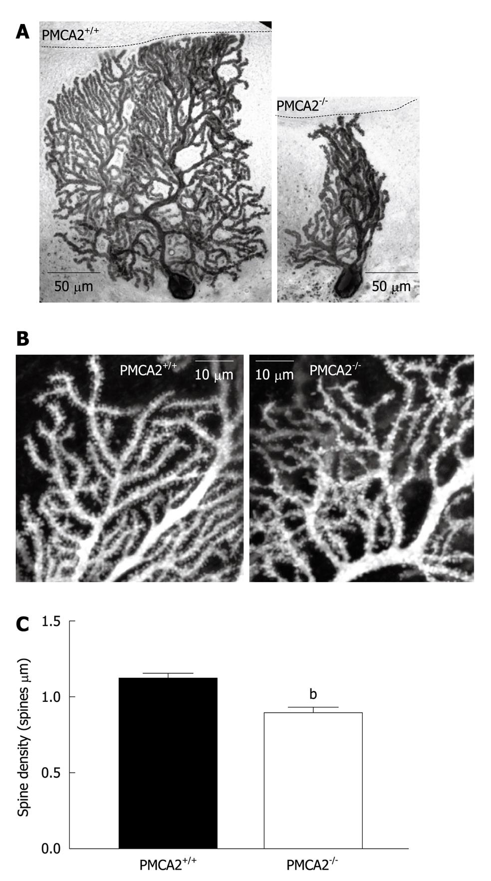
Figure 5 Dendritic morphology of PMCA2-/- PN dendrites is severely disrupted.
A: Representative PNs from wild type PMCA2+/+ (left) and PMCA2-/- (right) cerebellum. Note the reduced thickness of the ML in PMCA2-/- cerebellar cortex, as indicated by the black dashed line and the disorganized dendritic tree. PNs had been previously filled with biocytin during electrophysiological patch clamp recordings. Images shown here are after post-hoc identification of the cells with an anti-biocytin antibody; B: Higher resolution images show the tertiary dendrites in more detail from PNs collected in the same way as in (A). Images are reconstructed from a stack of 20 consecutive confocal slices at 0.1 μm intervals. Spines are visible along these tertiary dendrites in both examples, but the spines on the PMCA2-/- dendrites are less ordered and have lower density. C: The mean values from combined data from more than 90 segments of dendrite are shown, error bars are SE, bP < 0.001.
- Citation: Huang H, Nagaraja RY, Garside ML, Akemann W, Knöpfel T, Empson RM. Contribution of plasma membrane Ca2+ ATPase to cerebellar synapse function. World J Biol Chem 2010; 1(5): 95-102
- URL: https://www.wjgnet.com/1949-8454/full/v1/i5/95.htm
- DOI: https://dx.doi.org/10.4331/wjbc.v1.i5.95


