Copyright
©2010 Baishideng Publishing Group Co.
World J Biol Chem. May 26, 2010; 1(5): 109-132
Published online May 26, 2010. doi: 10.4331/wjbc.v1.i5.109
Published online May 26, 2010. doi: 10.4331/wjbc.v1.i5.109
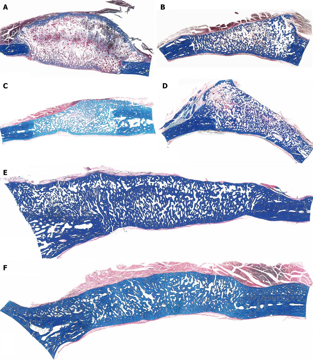
Figure 3 Calvarial tissue regeneration by doses of non-gamma irradiated recombinant human osteogenic protein-1 (hOP-1) implanted in non-healing calvarial defects of non-human primates P.
ursinus. A: Defect treated with 0.5 mg hOP-1 recombined with allogeneic insoluble collagenous bone matrix (ICBM) and harvested on day 30. Extensive mineralization and pronounced osteogenesis with displacement of the temporalis muscle overlying the implanted defect. Scattered remnants of the collagenous matrix as carrier embedded within a loose but highly vascular and cellular matrix; mineralized bone (in blue) facing the pericranial and endocranial aspect of the implanted hOP-1 osteogenic device; B: Remodeling and incorporation of the newly formed bone 90 d after implantation of 2.5 mg hOP-1 with corticalization of the endocranial aspect of the newly formed and mineralized bone; C, D: Exuberant induction of bone formation with peripheral corticalization of the newly formed bone 90 d after application of 0.1 and 2.5 mg hOP-1 per gram of allogeneic ICBM as carrier; E, F: Exuberant osteogenesis with solid block of remodeled bone particularly at the endocranial interface after implantation of 2.5 mg hOP-1 osteogenic devices harvested and processed for undecalcified histology on day 365 after implantation. A-F: Undecalcified sections cut at 6 μm stained free-floating with a modified Goldner’s trichrome.
- Citation: Ripamonti U. Soluble and insoluble signals sculpt osteogenesis in angiogenesis. World J Biol Chem 2010; 1(5): 109-132
- URL: https://www.wjgnet.com/1949-8454/full/v1/i5/109.htm
- DOI: https://dx.doi.org/10.4331/wjbc.v1.i5.109


