Copyright
©The Author(s) 2017.
World J Gastrointest Surg. Aug 27, 2017; 9(8): 182-185
Published online Aug 27, 2017. doi: 10.4240/wjgs.v9.i8.182
Published online Aug 27, 2017. doi: 10.4240/wjgs.v9.i8.182
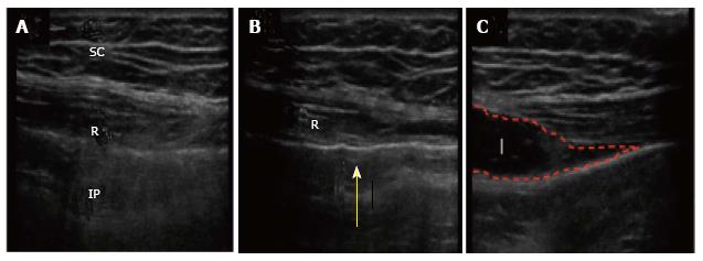
Figure 2 Ultrasound images.
A: The muscle layers; B: The Peti-needle™ positioned below the peritoneum, the notch was made by the needle tip (yellow arrow) before the needle was inserted via the peritoneum; C: Local analgesic was then administered into the correct layer. R: Rectus muscle; IP: Intraperitoneal space; SC: Subcutaneous tissue; I: Injectate.
- Citation: Nagata J, Watanabe J, Sawatsubashi Y, Akiyama M, Arase K, Minagawa N, Torigoe T, Hamada K, Nakayama Y, Hirata K. Novel technique of abdominal wall nerve block for laparoscopic colostomy: Rectus sheath block with transperitoneal approach. World J Gastrointest Surg 2017; 9(8): 182-185
- URL: https://www.wjgnet.com/1948-9366/full/v9/i8/182.htm
- DOI: https://dx.doi.org/10.4240/wjgs.v9.i8.182


