Copyright
©The Author(s) 2016.
World J Gastrointest Surg. Jun 27, 2016; 8(6): 467-471
Published online Jun 27, 2016. doi: 10.4240/wjgs.v8.i6.467
Published online Jun 27, 2016. doi: 10.4240/wjgs.v8.i6.467
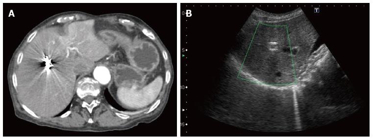
Figure 4 Post embolization imaging.
A: Transversal view on the contrast enhanced abdominal computed tomography performed two days after embolization showing that the coils are in place and the absence of blood extravasation; B: Thirty-days control liver ultrasound showing coils in place in the sagittal plane.
- Citation: Vultaggio F, Morère PH, Constantin C, Christodoulou M, Roulin D. Gastrointestinal bleeding and obstructive jaundice: Think of hepatic artery aneurysm. World J Gastrointest Surg 2016; 8(6): 467-471
- URL: https://www.wjgnet.com/1948-9366/full/v8/i6/467.htm
- DOI: https://dx.doi.org/10.4240/wjgs.v8.i6.467


