Copyright
©The Author(s) 2016.
World J Gastrointest Surg. Jun 27, 2016; 8(6): 467-471
Published online Jun 27, 2016. doi: 10.4240/wjgs.v8.i6.467
Published online Jun 27, 2016. doi: 10.4240/wjgs.v8.i6.467
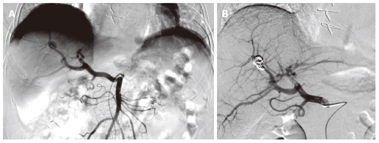
Figure 3 Supra-selective angiography of the hepatic artery branch aneurysm.
A: The left image is before embolization; it shows the hepatic artery splitting and the aneurysm taking source from one of the right branches; B: The right image is after supra-selective embolization of the aneurysm with microcoils. The catheter is still visible. Note that the hepatic artery originates from the superior mesenteric artery. This exhibits a full hepatic artery variant, type IX in Michels’ classification.
- Citation: Vultaggio F, Morère PH, Constantin C, Christodoulou M, Roulin D. Gastrointestinal bleeding and obstructive jaundice: Think of hepatic artery aneurysm. World J Gastrointest Surg 2016; 8(6): 467-471
- URL: https://www.wjgnet.com/1948-9366/full/v8/i6/467.htm
- DOI: https://dx.doi.org/10.4240/wjgs.v8.i6.467


