Copyright
©The Author(s) 2016.
World J Gastrointest Surg. Jun 27, 2016; 8(6): 467-471
Published online Jun 27, 2016. doi: 10.4240/wjgs.v8.i6.467
Published online Jun 27, 2016. doi: 10.4240/wjgs.v8.i6.467
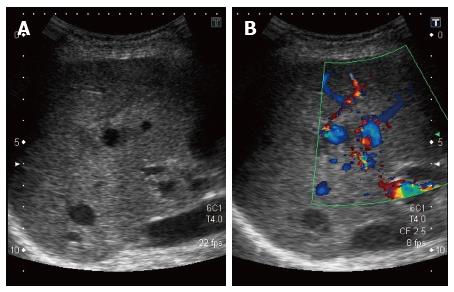
Figure 2 Aneurysm on hepatic Doppler-ultrasound.
Sagittal view of the liver representing the aneurysm at the center of the echographic field (A) and Doppler signals confirming the blood flow inside the aneurysm (B). The branch from the hepatic artery the aneurysm is originating from is not seen on the image.
- Citation: Vultaggio F, Morère PH, Constantin C, Christodoulou M, Roulin D. Gastrointestinal bleeding and obstructive jaundice: Think of hepatic artery aneurysm. World J Gastrointest Surg 2016; 8(6): 467-471
- URL: https://www.wjgnet.com/1948-9366/full/v8/i6/467.htm
- DOI: https://dx.doi.org/10.4240/wjgs.v8.i6.467


