
















©2014 Baishideng Publishing Group Inc.
World J Gastrointest Surg. Aug 27, 2014; 6(8): 160-163
Published online Aug 27, 2014. doi: 10.4240/wjgs.v6.i8.160
Published online Aug 27, 2014. doi: 10.4240/wjgs.v6.i8.160
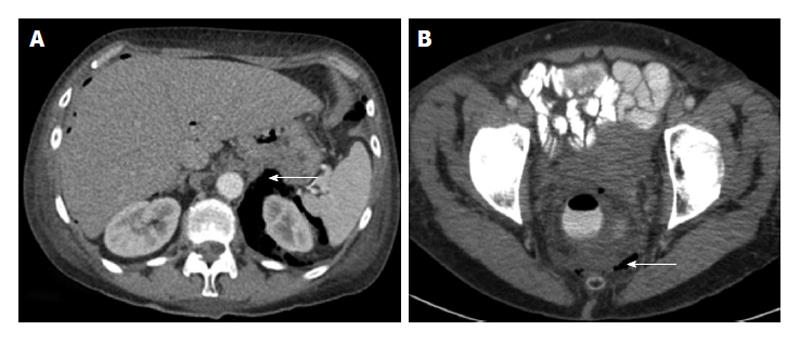
Figure 3 Computed tomography-scan.
A: Abdominal computed tomography-scan showing retroperitoneal air surrounding the left kidney (arrow); B: Pelvic computed tomography-scan with rectal contrast showing free air in the mesorectum (arrow).
- Citation: Simkens GA, Nienhuijs SW, Luyer MD, Hingh IH. Massive surgical emphysema following transanal endoscopic microsurgery. World J Gastrointest Surg 2014; 6(8): 160-163
- URL: https://www.wjgnet.com/1948-9366/full/v6/i8/160.htm
- DOI: https://dx.doi.org/10.4240/wjgs.v6.i8.160