
















©2014 Baishideng Publishing Group Co.
World J Gastrointest Surg. Apr 27, 2014; 6(4): 65-69
Published online Apr 27, 2014. doi: 10.4240/wjgs.v6.i4.65
Published online Apr 27, 2014. doi: 10.4240/wjgs.v6.i4.65
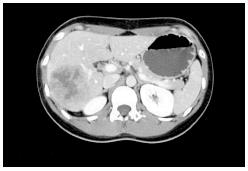
Figure 1 A computed tomography scan with nonionic contrast confirmed a mass located in the posterior right lobe within segments VI-VII and measuring 7.
2 cm × 6.0 cm. The lesion demonstrated peripheral enhancement with central necrosis but no evidence for portal vein invasion. The hepatic veins were patent and no biliary dilatation was observed. No pulmonary lesion was highlighted.
- Citation: Labgaa I, Carrasco-Avino G, Fiel MI, Schwartz ME. Pancreatic recurrence of intrahepatic cholangiocarcinoma: Case report and review of the literature. World J Gastrointest Surg 2014; 6(4): 65-69
- URL: https://www.wjgnet.com/1948-9366/full/v6/i4/65.htm
- DOI: https://dx.doi.org/10.4240/wjgs.v6.i4.65