Copyright
©2014 Baishideng Publishing Group Inc.
World J Gastrointest Surg. Oct 27, 2014; 6(10): 190-200
Published online Oct 27, 2014. doi: 10.4240/wjgs.v6.i10.190
Published online Oct 27, 2014. doi: 10.4240/wjgs.v6.i10.190
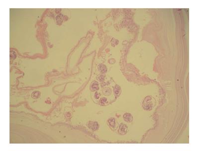
Figure 3 Cyst wall of the patient’s mass consists of a laminated faintly stained chitinous membrane (outer layer).
Multiple protoscolices are present within the daughter cyst (inner germinal layer, hematoxylin-Eosin stain × 100).
- Citation: Akbulut S, Yavuz R, Sogutcu N, Kaya B, Hatipoglu S, Senol A, Demircan F. Hydatid cyst of the pancreas: Report of an undiagnosed case of pancreatic hydatid cyst and brief literature review. World J Gastrointest Surg 2014; 6(10): 190-200
- URL: https://www.wjgnet.com/1948-9366/full/v6/i10/190.htm
- DOI: https://dx.doi.org/10.4240/wjgs.v6.i10.190


