Copyright
©2014 Baishideng Publishing Group Inc.
World J Gastrointest Surg. Oct 27, 2014; 6(10): 190-200
Published online Oct 27, 2014. doi: 10.4240/wjgs.v6.i10.190
Published online Oct 27, 2014. doi: 10.4240/wjgs.v6.i10.190
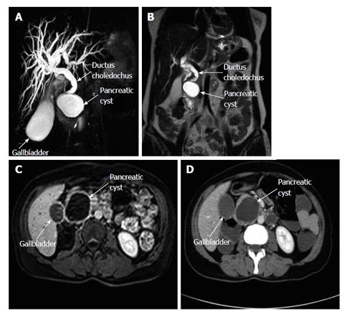
Figure 1 Magnetic resonance cholangiopancreatography shows a hydropic gall bladder and dilated intrahepatic bile ducts and common bile duct.
The distal portion of the common bile duct is narrowed due to external compression. A: A pancreatic cyst compressing the lower tip of the common bile duct is also seen in this section; B: A coronal T2-weighed MR cross-section shows a pancreatic cyst and a dilated common bile duct; C: Intravenous T1-weighed axial cross-section with contrast enhancement shows a pancreatic cyst; D: An axial computerized tomography cross-section with contrast enhancement shows a pancreatic cyst with a thick wall and without central contrast uptake. MR: Magnetic resonance.
- Citation: Akbulut S, Yavuz R, Sogutcu N, Kaya B, Hatipoglu S, Senol A, Demircan F. Hydatid cyst of the pancreas: Report of an undiagnosed case of pancreatic hydatid cyst and brief literature review. World J Gastrointest Surg 2014; 6(10): 190-200
- URL: https://www.wjgnet.com/1948-9366/full/v6/i10/190.htm
- DOI: https://dx.doi.org/10.4240/wjgs.v6.i10.190


