Copyright
©2013 Baishideng Publishing Group Co.
World J Gastrointest Surg. Apr 27, 2013; 5(4): 83-96
Published online Apr 27, 2013. doi: 10.4240/wjgs.v5.i4.83
Published online Apr 27, 2013. doi: 10.4240/wjgs.v5.i4.83
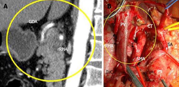
Figure 7 In 75-year-old woman (case 1), 360° PDAC encasement of the replaced right hepatic artery was diagnosed on computed tomography (A), while endoUS data described only tumor abutment with the artery.
Arterial phase, Sagittal images: CT showed circumferential infiltration of the rRHA, B: Intraoperative photograph. An extended Whipple procedure was performed. There were no signs of rRHA or SMA involvement during surgery (arrows). The level of resection was R1 because of the contact of the rRHA with the tumor. SMA: Superior mesenteric artery; rRHA: Replaced right hepatic; LHA: Left hepatic; RGEA: Right gastro-epiploic arteries; CT: Celiac trunk; SMV: Superior mesenteric; PV: Portal; LRV: Left renal vein; T: Tumor; PS: Pancreatic stump.
- Citation: Egorov VI, Petrov RV, Solodinina EN, Karmazanovsky GG, Starostina NS, Kuruschkina NA. Computed tomography-based diagnostics might be insufficient in the determination of pancreatic cancer unresectability. World J Gastrointest Surg 2013; 5(4): 83-96
- URL: https://www.wjgnet.com/1948-9366/full/v5/i4/83.htm
- DOI: https://dx.doi.org/10.4240/wjgs.v5.i4.83


