Copyright
©2010 Baishideng.
World J Gastrointest Surg. Apr 27, 2010; 2(4): 147-152
Published online Apr 27, 2010. doi: 10.4240/wjgs.v2.i4.147
Published online Apr 27, 2010. doi: 10.4240/wjgs.v2.i4.147
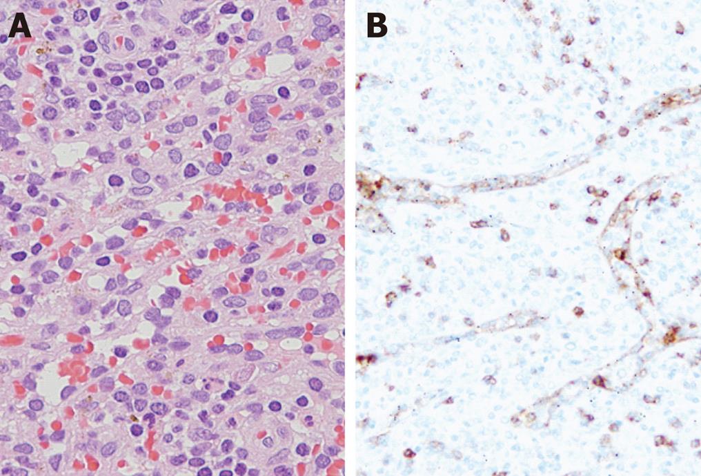
Figure 4 Pathology of the splenic lesion.
A: Microscopically, the mass is composed of predominantly a red pulp splenic sinusoidal structure accompanied by lymphocytes and macrophages. Atypical cells or mitosis were not observed; B: The endothelial cells lining the sinusoidal structure are immunoreactive for CD8.
- Citation: Namikawa T, Kitagawa H, Iwabu J, Kobayashi M, Matsumoto M, Hanazaki K. Laparoscopic splenectomy for splenic hamartoma: Case management and clinical consequences. World J Gastrointest Surg 2010; 2(4): 147-152
- URL: https://www.wjgnet.com/1948-9366/full/v2/i4/147.htm
- DOI: https://dx.doi.org/10.4240/wjgs.v2.i4.147


