
















©2010 Baishideng Publishing Group Co.
World J Gastrointest Surg. Dec 27, 2010; 2(12): 405-408
Published online Dec 27, 2010. doi: 10.4240/wjgs.v2.i12.405
Published online Dec 27, 2010. doi: 10.4240/wjgs.v2.i12.405
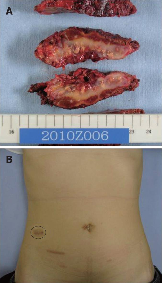
Figure 5 Resected specimen (A) and postoperative picture of abdomen (B) of case 2.
A: Resected specimen revealed suppurative inflammatory tissue which consisted of abscess, granulomatous- and fibrous tissue in and around the liver; B: Circle indicates surgical wound of single port. Surgical scars of previous appendectomy are seen.
- Citation: Hayashi M, Asakuma M, Tsunemi S, Inoue Y, Shimizu T, Komeda K, Hirokawa F, Takeshita A, Egashira Y, Tanigawa N. Surgical treatment for abdominal actinomycosis: A report of two cases. World J Gastrointest Surg 2010; 2(12): 405-408
- URL: https://www.wjgnet.com/1948-9366/full/v2/i12/405.htm
- DOI: https://dx.doi.org/10.4240/wjgs.v2.i12.405