Copyright
©2010 Baishideng Publishing Group Co.
World J Gastrointest Surg. Oct 27, 2010; 2(10): 331-336
Published online Oct 27, 2010. doi: 10.4240/wjgs.v2.i10.331
Published online Oct 27, 2010. doi: 10.4240/wjgs.v2.i10.331
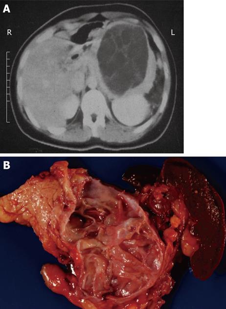
Figure 1 Typical computed tomography (A) and gross (B) appearance of a mucinous cystic neoplasm showing the distal location and the lack of communication with the duct, respectively.
- Citation: Cunningham SC, Hruban RH, Schulick RD. Differentiating intraductal papillary mucinous neoplasms from other pancreatic cystic lesions. World J Gastrointest Surg 2010; 2(10): 331-336
- URL: https://www.wjgnet.com/1948-9366/full/v2/i10/331.htm
- DOI: https://dx.doi.org/10.4240/wjgs.v2.i10.331


