Copyright
©2010 Baishideng Publishing Group Co.
World J Gastrointest Surg. Oct 27, 2010; 2(10): 324-330
Published online Oct 27, 2010. doi: 10.4240/wjgs.v2.i10.324
Published online Oct 27, 2010. doi: 10.4240/wjgs.v2.i10.324
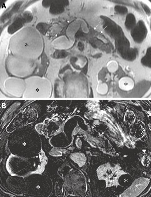
Figure 2 Axial T2-weighted (A) and subtraction (post-contrast minus pre-contrast) (B) images at the level of the pancreas demonstrate marked enlargement of the main pancreatic duct (arrowheads) with intraluminal enhancing papillary projections (arrows).
Main duct intraductal papillary mucinous neoplasm with in situ carcinoma was confirmed at histopathology after total pancreatectomy. Multiple renal cysts (asterisks).
- Citation: Pedrosa I, Boparai D. Imaging considerations in intraductal papillary mucinous neoplasms of the pancreas. World J Gastrointest Surg 2010; 2(10): 324-330
- URL: https://www.wjgnet.com/1948-9366/full/v2/i10/324.htm
- DOI: https://dx.doi.org/10.4240/wjgs.v2.i10.324


