
















Copyright: ©Author(s) 2026.
World J Gastrointest Surg. Sep 27, 2026; 18(9): 123459
Published online Sep 27, 2026. doi: 10.4240/wjgs.123459
Published online Sep 27, 2026. doi: 10.4240/wjgs.123459
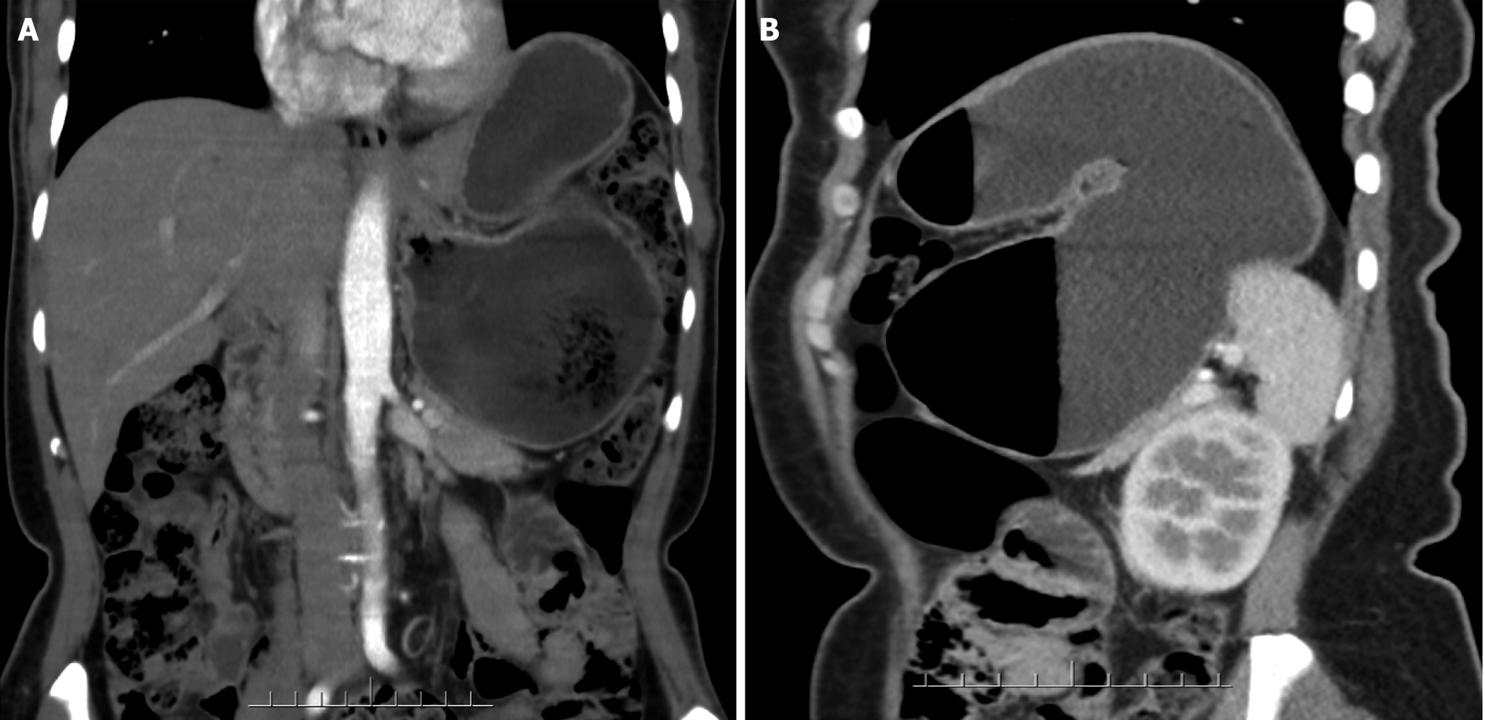
Figure 1 Contrast-enhanced abdominal computed tomography.
A: The coronal view, it shows elevation of the left side of the diaphragm, upward displa
- Citation: Wang S, Yang XF, Wang W. Laparoscopic reduction of gastric volvulus: A case report and review of literature. World J Gastrointest Surg 2026; 18(9): 123459
- URL: https://www.wjgnet.com/1948-9366/full/v18/i9/123459.htm
- DOI: https://dx.doi.org/10.4240/wjgs.123459