
















Copyright: ©Author(s) 2026.
World J Gastrointest Surg. Sep 27, 2026; 18(9): 122838
Published online Sep 27, 2026. doi: 10.4240/wjgs.122838
Published online Sep 27, 2026. doi: 10.4240/wjgs.122838
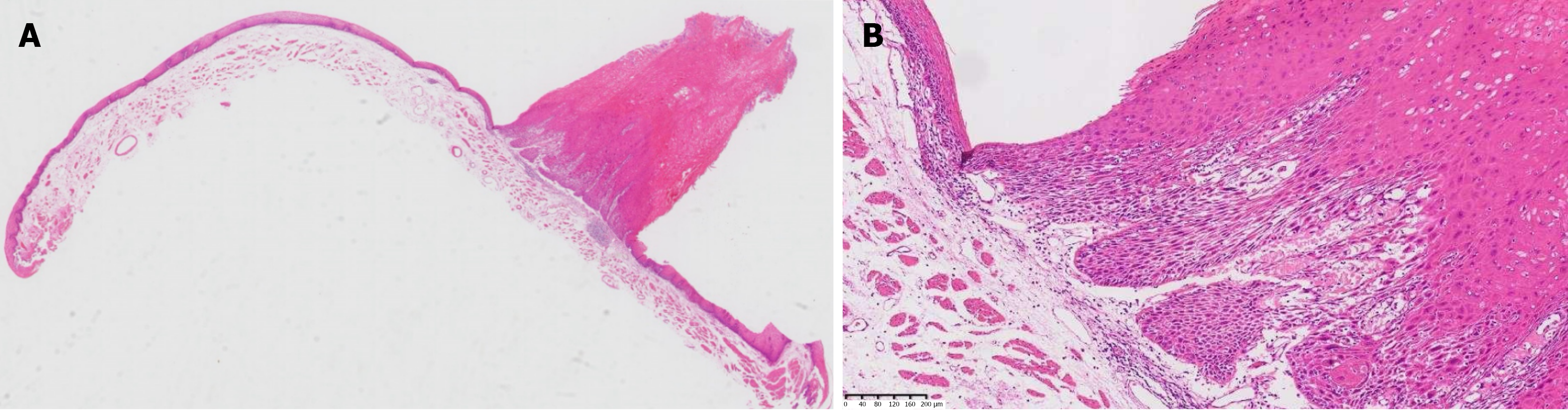
Figure 4 Histopathological features of the esophageal endoscopic submucosal dissection specimen (hematoxylin and eosin staining).
A: Esophageal endoscopic submucosal dissection (ESD) specimen [4 × hematoxylin and eosin (HE)] showing an esophageal mass displaying carcinoma in situ (verrucous or papillary) with a stone pillar-like appearance 31 cm from the incisors; B: Esophageal ESD specimen (10 × HE) showing epithelial cell dysplasia, with no infiltration of the basal cells by the carcinoma in situ.
- Citation: Zhang X, Zhou JY, Wu ZW, Zhou SX, Zheng JF, Zheng WY, Pan WS, Wu HG. Coexistence of esophageal verrucous carcinoma in situ and gastric intramucosal poorly differentiated adenocarcinoma: A case report. World J Gastrointest Surg 2026; 18(9): 122838
- URL: https://www.wjgnet.com/1948-9366/full/v18/i9/122838.htm
- DOI: https://dx.doi.org/10.4240/wjgs.122838