
















Copyright: ©Author(s) 2026.
World J Gastrointest Surg. Sep 27, 2026; 18(9): 122838
Published online Sep 27, 2026. doi: 10.4240/wjgs.122838
Published online Sep 27, 2026. doi: 10.4240/wjgs.122838
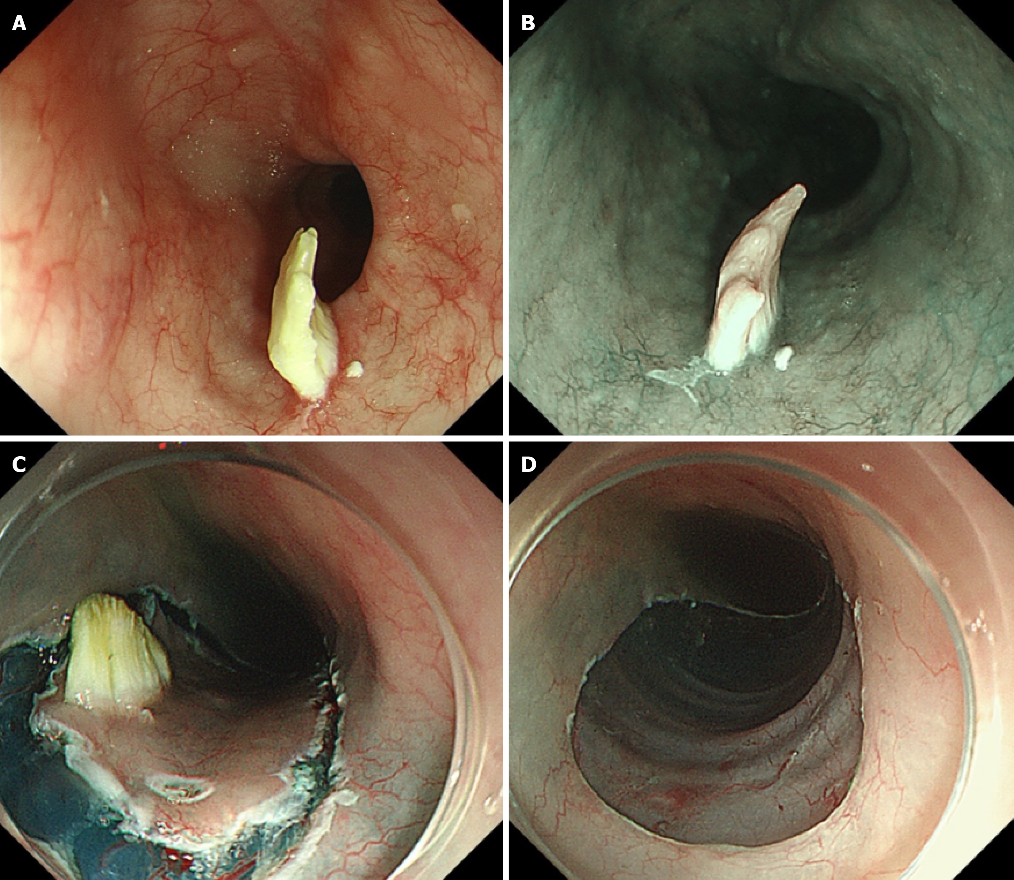
Figure 3 Endoscopic manifestations of the progressive esophageal lesion and the endoscopic submucosal dissection procedure.
A: The lesion appears more protruded than before, showing a whitish and slightly yellowish appearance under white light; B: Narrow-band imaging revealed irregular blood vessels, while the surface structure remained indiscernible; C and D: Endoscopic submucosal dissection was performed to completely excise the lesion.
- Citation: Zhang X, Zhou JY, Wu ZW, Zhou SX, Zheng JF, Zheng WY, Pan WS, Wu HG. Coexistence of esophageal verrucous carcinoma in situ and gastric intramucosal poorly differentiated adenocarcinoma: A case report. World J Gastrointest Surg 2026; 18(9): 122838
- URL: https://www.wjgnet.com/1948-9366/full/v18/i9/122838.htm
- DOI: https://dx.doi.org/10.4240/wjgs.122838