
















Copyright: ©Author(s) 2026.
World J Gastrointest Surg. Sep 27, 2026; 18(9): 122761
Published online Sep 27, 2026. doi: 10.4240/wjgs.122761
Published online Sep 27, 2026. doi: 10.4240/wjgs.122761
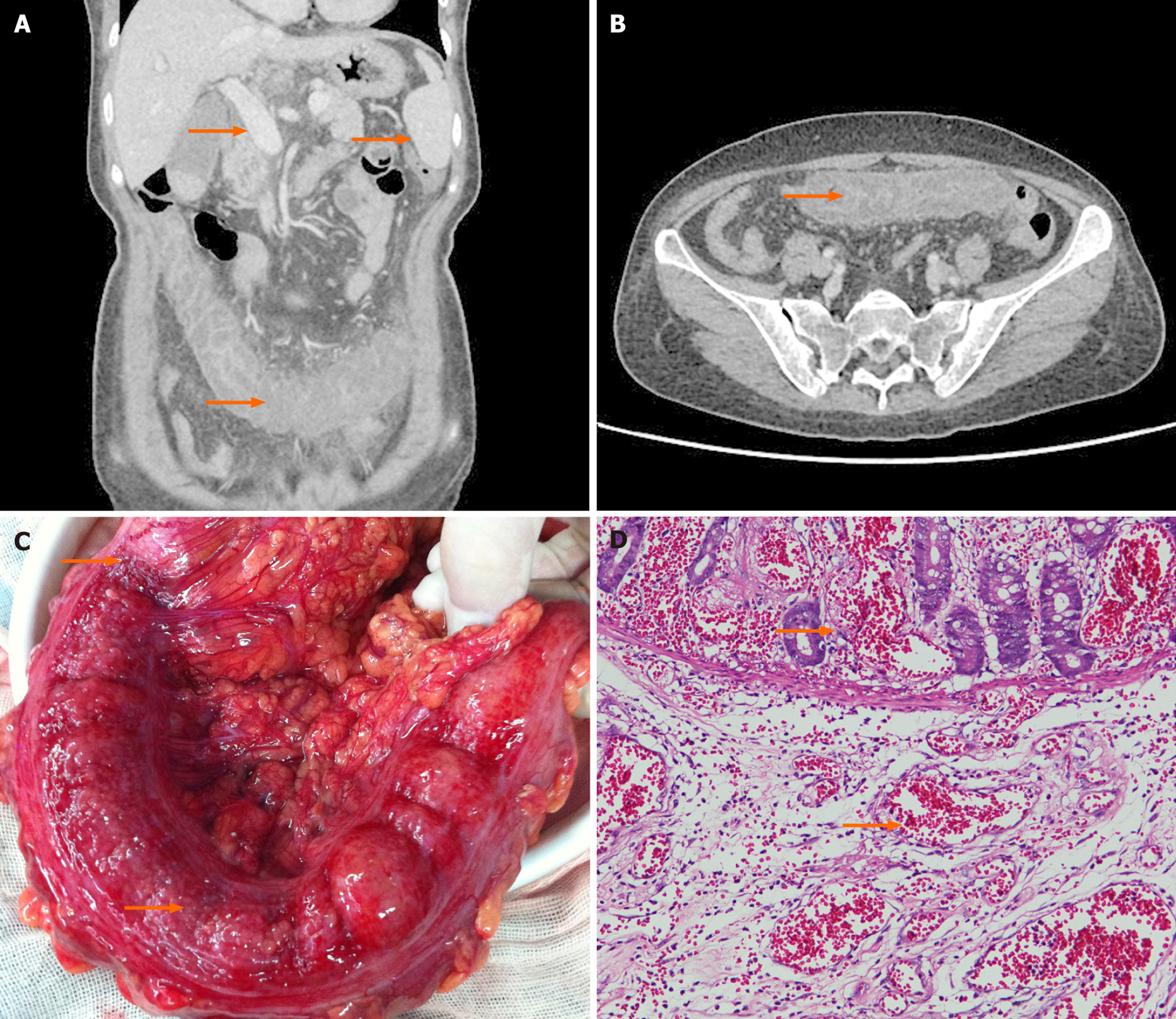
Figure 2 Imaging, intraoperative and histopathological findings.
A: Contrast-enhanced abdominal computed tomography revealed portal hypertension, splenomegaly, tortuous venous clusters at the splenic hilum, and hemorrhage and edema of the transverse colon (orange arrows); B: Computed tomography revealed marked thickening and narrowing of the transverse colon compared with adjacent bowel loops (orange arrow); C: Intraoperative examination revealed a dark red transverse colon, with a subserosal hematoma-like change and clear demarcation from the non-congested colon (orange arrows); D: Histopathological analysis (hematoxylin and eosin stain) showed marked vascular congestion with hemorrhage, few intraluminal thrombi, focal mucosal necrosis and shedding, marked submucosal edema with vascular proliferation, thick-walled vessels, and focal angioma-like hyperplasia (orange arrows).
- Citation: Ge JK, Tian HB, Zou HB, He LY. Essential thrombocythemia presenting as localized hemorrhage and mucosal necrosis of transverse colon: A case report. World J Gastrointest Surg 2026; 18(9): 122761
- URL: https://www.wjgnet.com/1948-9366/full/v18/i9/122761.htm
- DOI: https://dx.doi.org/10.4240/wjgs.122761