
















Copyright: ©Author(s) 2026.
World J Gastrointest Surg. Sep 27, 2026; 18(9): 121842
Published online Sep 27, 2026. doi: 10.4240/wjgs.121842
Published online Sep 27, 2026. doi: 10.4240/wjgs.121842
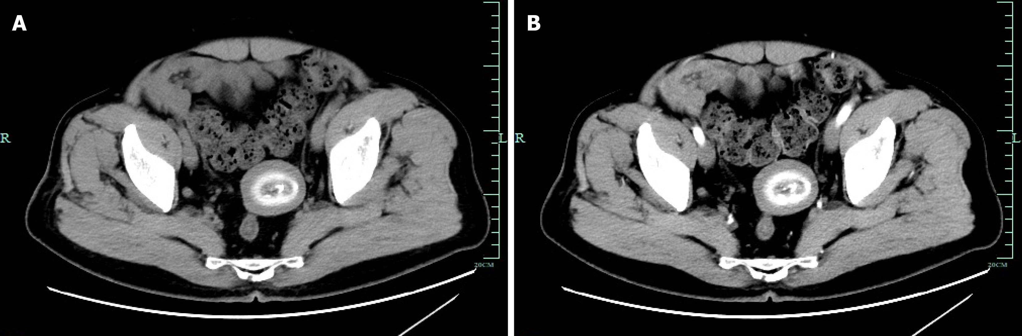
Figure 1 Abdominal computed tomography.
A: Plain abdominal computed tomography (CT) shows a 5.0 cm × 4.2 cm cystic-solid mass within the left pelvic margin with well-defined borders; B: Contrast-enhanced abdominal CT shows no obvious enhancement or significant blood supply within the mass.
- Citation: Lou YM, Gao TZ, Zheng CZ, Yu H, Hong YY, Liu XS. Microscopic evaluation of giant peritoneal loose body: A case report. World J Gastrointest Surg 2026; 18(9): 121842
- URL: https://www.wjgnet.com/1948-9366/full/v18/i9/121842.htm
- DOI: https://dx.doi.org/10.4240/wjgs.121842