
















Copyright: ©Author(s) 2026.
World J Gastrointest Surg. Sep 27, 2026; 18(9): 119675
Published online Sep 27, 2026. doi: 10.4240/wjgs.119675
Published online Sep 27, 2026. doi: 10.4240/wjgs.119675
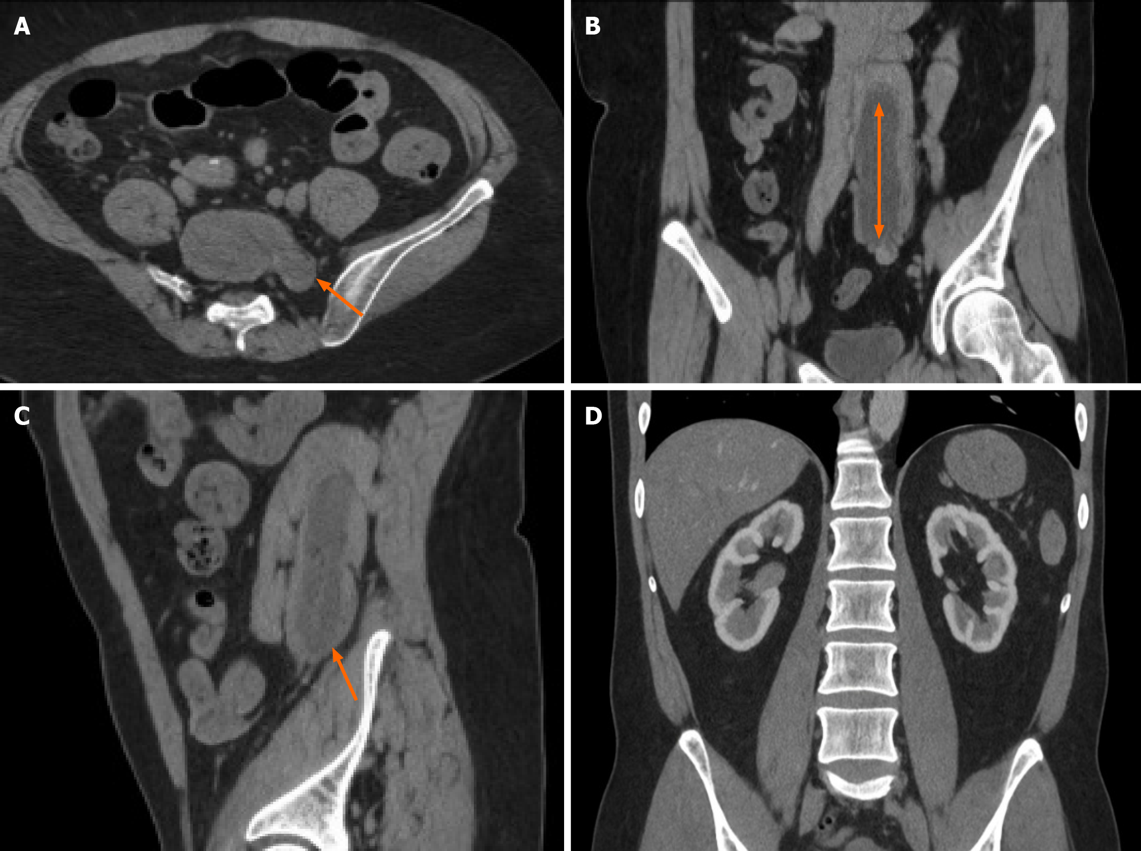
Figure 5 Typical preoperative computed tomography imaging findings in a patient from the study cohort.
These images illustrate the common tumor characteristics integral to the prediction models. A: Axial contrast-enhanced computed tomography image showing irregular sigmoid colon wall thickening (arrow) with a maximum thickness of approximately 8.2 mm, consistent with a T3 stage tumor (penetrating the muscularis propria); B: Coronal reconstructed image demonstrating the craniocaudal extent of the tumor measuring approximately 4.6 cm (double-headed arrow); C: The sagittal image clearly depicts the relationship between the tumor and adjacent structures; the presence of preserved fat planes suggests no invasion of the surrounding organs; D: Coronal enhancement image showing homogeneous enhancement of the tumor with no evidence of distant metastasis. These features (T stage and tumor size) were key variables in the multivariate analysis.
- Citation: Qiang JH, Cheng J, Meng F, Zhang SQ. Application of multivariate logistic regression in predicting postoperative intestinal function recovery and lymph node metastasis in colorectal cancer. World J Gastrointest Surg 2026; 18(9): 119675
- URL: https://www.wjgnet.com/1948-9366/full/v18/i9/119675.htm
- DOI: https://dx.doi.org/10.4240/wjgs.119675