
















Copyright: ©Author(s) 2026.
World J Gastrointest Surg. Aug 27, 2026; 18(8): 121170
Published online Aug 27, 2026. doi: 10.4240/wjgs.121170
Published online Aug 27, 2026. doi: 10.4240/wjgs.121170
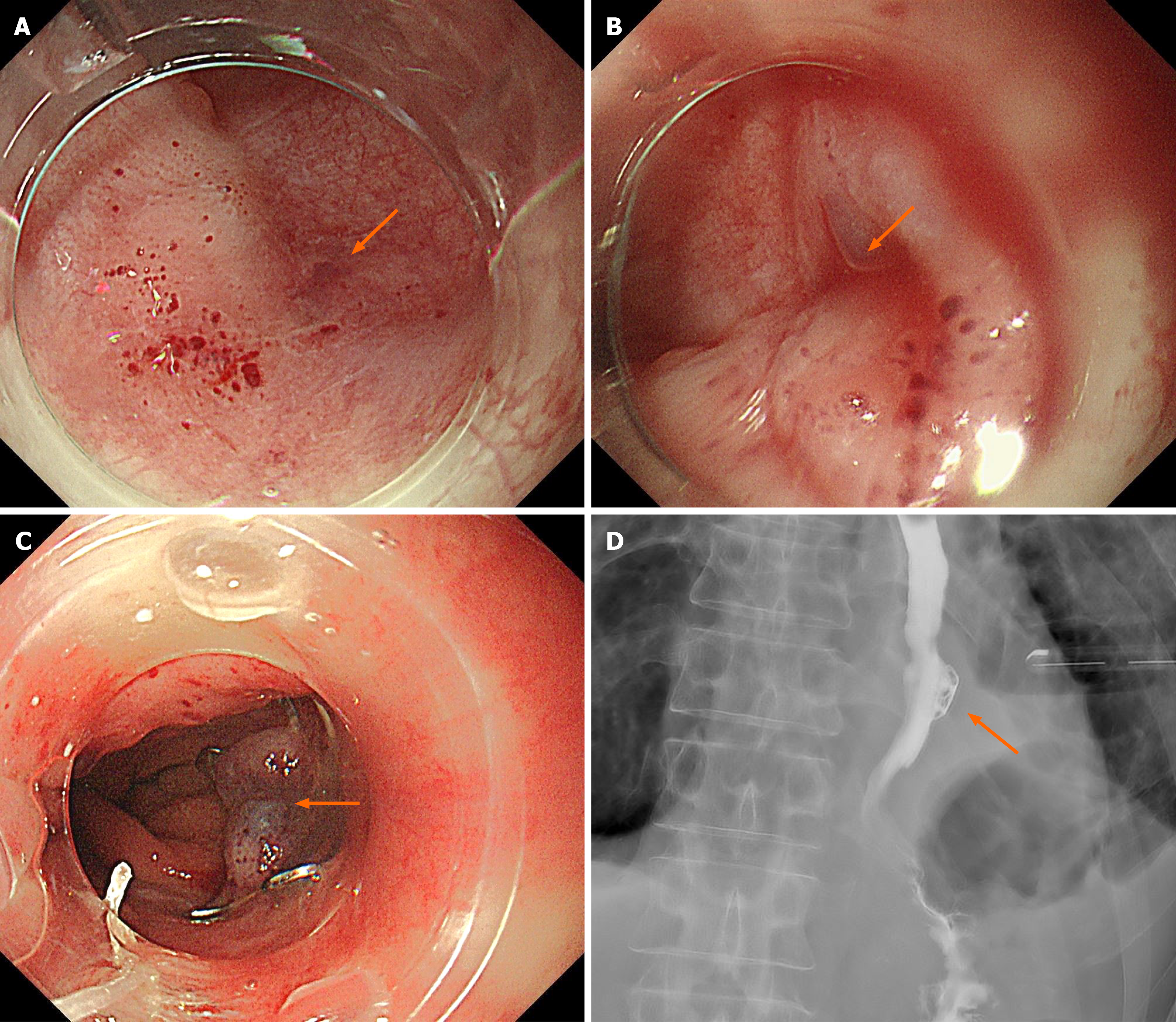
Figure 4 Endoscopic closure using an over-the-scope clip.
A: Endoscopic view of the 2-mm fistula located on the left wall just above the esophagogastric junction; B: The fistula and surrounding mucosa were suctioned into the over-the-scope clip (OTSC) cap to achieve adequate tissue capture; C: The OTSC was deployed after confirmation of sufficient tissue inversion into the cap, achieving full-thickness closure; D: Immediate water-soluble esophagography after OTSC placement demonstrated no visualization of the fistula and no leakage into the left pleural cavity; additional clips were not required; the esophagopleural fistula is indicated by an arrow.
- Citation: Takahashi N, Baba S, Toya Y, Nikai H, Fujisawa R, Endo F, Umemura A, Iwaya T, Nitta H, Sasaki A. Curative endoscopic closure of a chronic esophagopleural fistula after Boerhaave syndrome using an over-the-scope clip: A case report. World J Gastrointest Surg 2026; 18(8): 121170
- URL: https://www.wjgnet.com/1948-9366/full/v18/i8/121170.htm
- DOI: https://dx.doi.org/10.4240/wjgs.121170