
















Copyright: ©Author(s) 2026.
World J Gastrointest Surg. Aug 27, 2026; 18(8): 121170
Published online Aug 27, 2026. doi: 10.4240/wjgs.121170
Published online Aug 27, 2026. doi: 10.4240/wjgs.121170
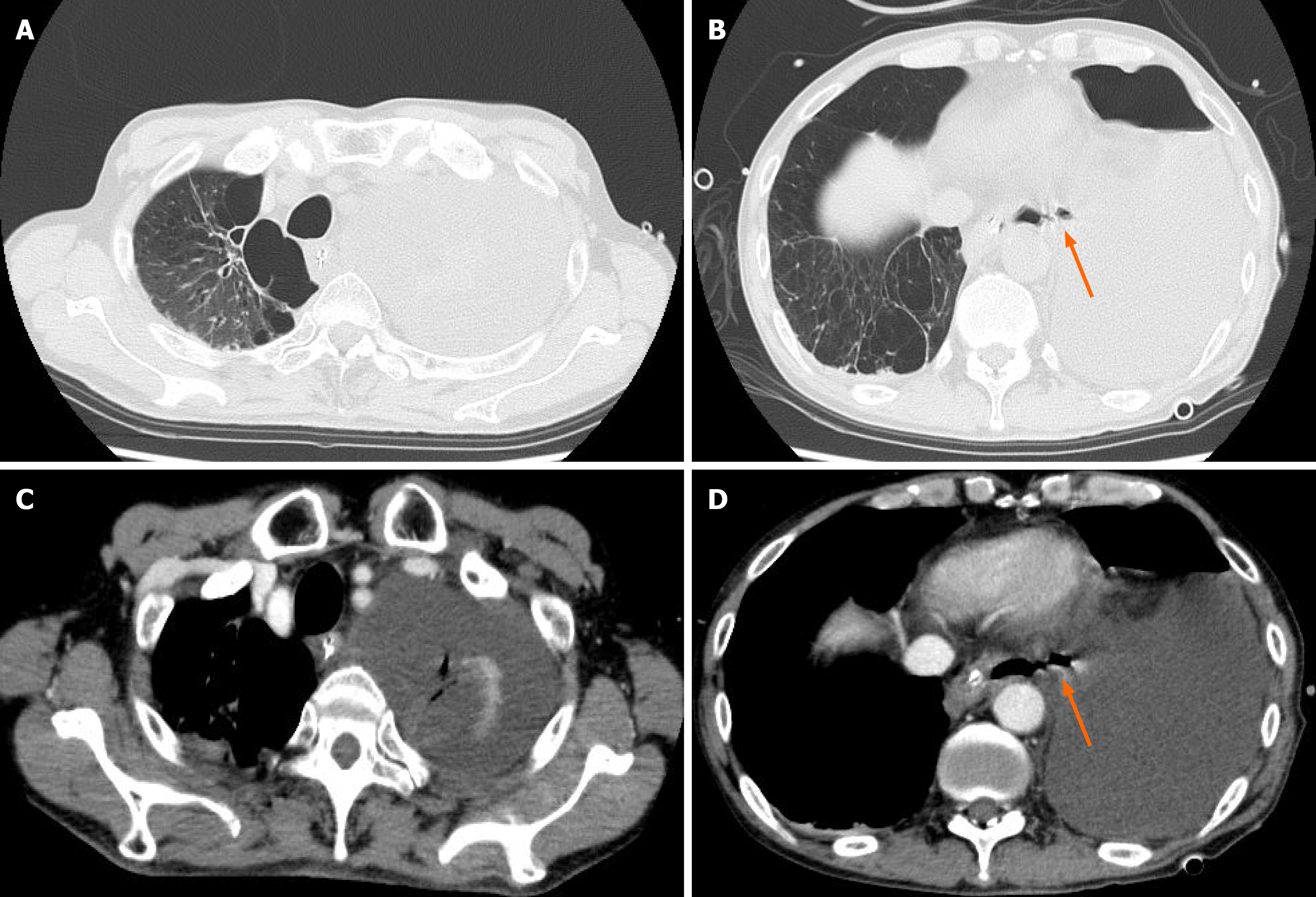
Figure 1 Contrast-enhanced computed tomography findings at the onset of spontaneous esophageal rupture.
A: A large amount of fluid was collected in the left thoracic cavity with left-lung collapse and rightward mediastinal shift, and a nasogastric tube for decompression was in place; B: Continuity between the lower esophageal lumen and the left pleural cavity suggestive of esophageal perforation; the ruptured esophageal wall is indicated by an arrow. No mediastinal or subcutaneous emphysema was observed; C: Extensive fluid was collected in the left thoracic cavity, with compression of the left lung; D: Suspected fistulous communication between the lower esophagus and the left pleural cavity; the ruptured esophageal wall is indicated by an arrow. Fluid accumulation with an air–fluid level was present in the left thoracic cavity.
- Citation: Takahashi N, Baba S, Toya Y, Nikai H, Fujisawa R, Endo F, Umemura A, Iwaya T, Nitta H, Sasaki A. Curative endoscopic closure of a chronic esophagopleural fistula after Boerhaave syndrome using an over-the-scope clip: A case report. World J Gastrointest Surg 2026; 18(8): 121170
- URL: https://www.wjgnet.com/1948-9366/full/v18/i8/121170.htm
- DOI: https://dx.doi.org/10.4240/wjgs.121170