
















Copyright: ©Author(s) 2026.
World J Gastrointest Surg. Aug 27, 2026; 18(8): 121137
Published online Aug 27, 2026. doi: 10.4240/wjgs.121137
Published online Aug 27, 2026. doi: 10.4240/wjgs.121137
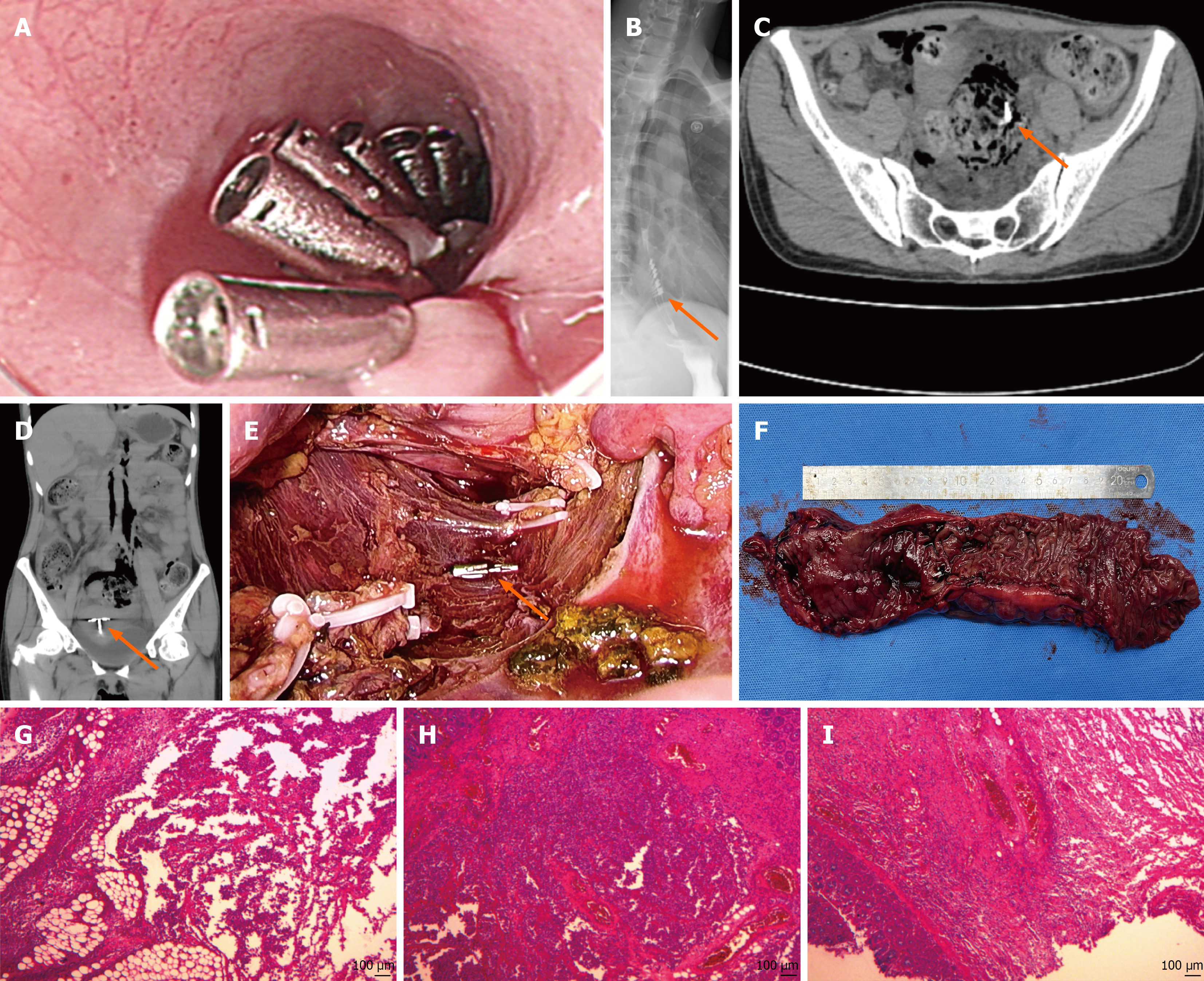
Figure 1 Imaging results.
A: Metallic clips were used to close the mucosal layer after peroral endoscopic myotomy surgery; B: Upper gastrointestinal contrast study was performed on postoperative day 3 after peroral endoscopic myotomy, revealing several metallic clip shadows (arrow) in the distal esophagus; C: Abdominal computed tomography scan showed a metal clip was identified adjacent to the perforation site (arrow); D: Coronal computed tomography image clearly depicted the clip (arrow); E: Laparoscopic exploration showed a metal clip was identified adjacent to the perforation site (arrow); F: Resected sigmoid specimen; G: Submucosal abscess with mixed neutrophilic and lymphocytic infiltration (intestinal wall); H: Submucosal abscess with mixed inflammation, necrosis, and focal vascular congestion (intestinal wall); I: Fibroadipose tissue with abscess, mixed inflammation, and focal vascular dilation/hemorrhage (mesentery).
- Citation: Zhang GY, Chen SG, Gao ZT, Huang ZT, Che SF, Li L, Long B, Ding FF, Yu ZY, Zhou HN, Jiao ZY. Metallic clip-associated colonic perforation after peroral endoscopic myotomy: A case report. World J Gastrointest Surg 2026; 18(8): 121137
- URL: https://www.wjgnet.com/1948-9366/full/v18/i8/121137.htm
- DOI: https://dx.doi.org/10.4240/wjgs.121137