
















Copyright: ©Author(s) 2026.
World J Gastrointest Surg. Aug 27, 2026; 18(8): 120911
Published online Aug 27, 2026. doi: 10.4240/wjgs.120911
Published online Aug 27, 2026. doi: 10.4240/wjgs.120911
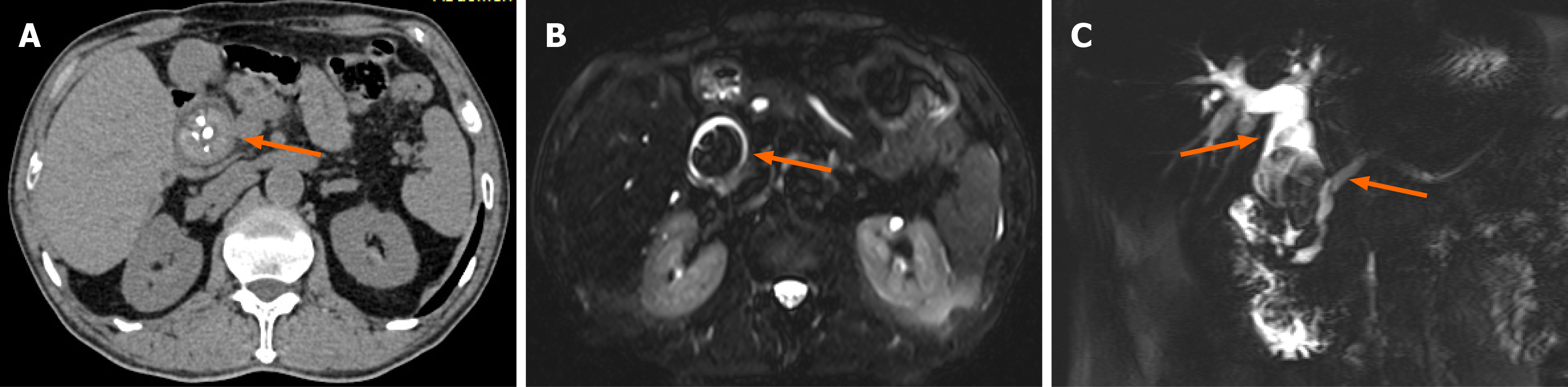
Figure 1 Computed tomography and magnetic resonance imaging findings.
A: Axial computed tomography image showing a large common bile duct (CBD) stone; B: Axial magnetic resonance imaging image confirming the presence of a large CBD stone; C: Magnetic resonance cholangiopancreatography indicating a large CBD stone with marked dilation of intrahepatic and extrahepatic bile ducts (left arrow) and dilation of the pancreatic duct (right arrow).
- Citation: Wang WF, Qiao ZY, Li Z, Zheng KZ, Sun DQ, Sheng MH. Endoscopic management of common bile duct stump fistula after choledochojejunostomy: A case report. World J Gastrointest Surg 2026; 18(8): 120911
- URL: https://www.wjgnet.com/1948-9366/full/v18/i8/120911.htm
- DOI: https://dx.doi.org/10.4240/wjgs.120911