
















Copyright: ©Author(s) 2026.
World J Gastrointest Surg. Aug 27, 2026; 18(8): 120360
Published online Aug 27, 2026. doi: 10.4240/wjgs.120360
Published online Aug 27, 2026. doi: 10.4240/wjgs.120360
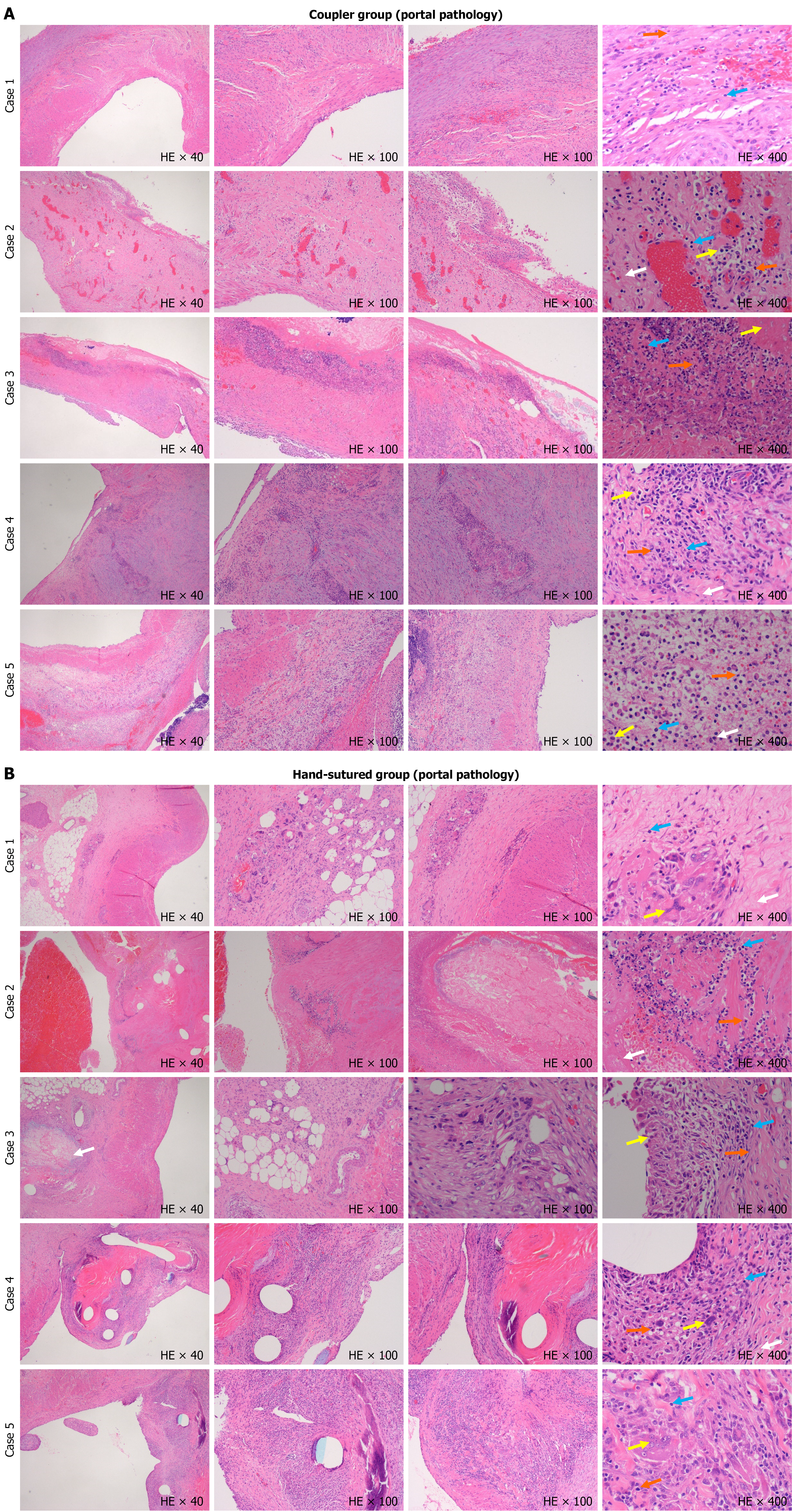
Figure 5 Histological analysis of portal vein tissue in the coupler group/hand-sutured group.
A: Histological analysis of portal vein tissue in the coupler group (hematoxylin and eosin staining). Representative images from five cases at 40 ×, 100 ×, and 400 ×. 40 ×/100 ×: The portal vein exhibits intact structure with minimal inflammation and no significant necrosis. Vessel walls are well preserved. 400 ×: Scattered macrophages (yellow arrows), lymphocytes (blue arrows), and rare plasma cells (orange arrows) are seen, with only small necrotic areas (white arrows); B: Histological analysis of portal vein tissue in the hand-sutured group (hematoxylin and eosin staining). Representative images from five cases at 40 ×, 100 ×, and 400 ×. 40 ×/100 ×: Structural disorganization, inflammatory cell infiltration, and focal necrosis are evident. Vessel walls show irregular thickening and fibrosis. 400 ×: Increased macrophages (yellow arrows), lymphocytes (blue arrows), plasma cells (orange arrows), and significant necrotic tissue (white arrows) are present, indicating higher inflammation and tissue injury.
- Citation: Li JJ, Yang J, Xie Y, Shang JM, Li J, Chen CY, Liu LN, Zhang ZY, Pan BH, Jiang WT. Sutureless portal vein reconstruction in porcine orthotopic liver transplantation using a microvascular coupler: A randomized study with artificial intelligence-assisted histopathology. World J Gastrointest Surg 2026; 18(8): 120360
- URL: https://www.wjgnet.com/1948-9366/full/v18/i8/120360.htm
- DOI: https://dx.doi.org/10.4240/wjgs.120360