
















Copyright: ©Author(s) 2026.
World J Gastrointest Surg. Aug 27, 2026; 18(8): 120285
Published online Aug 27, 2026. doi: 10.4240/wjgs.120285
Published online Aug 27, 2026. doi: 10.4240/wjgs.120285
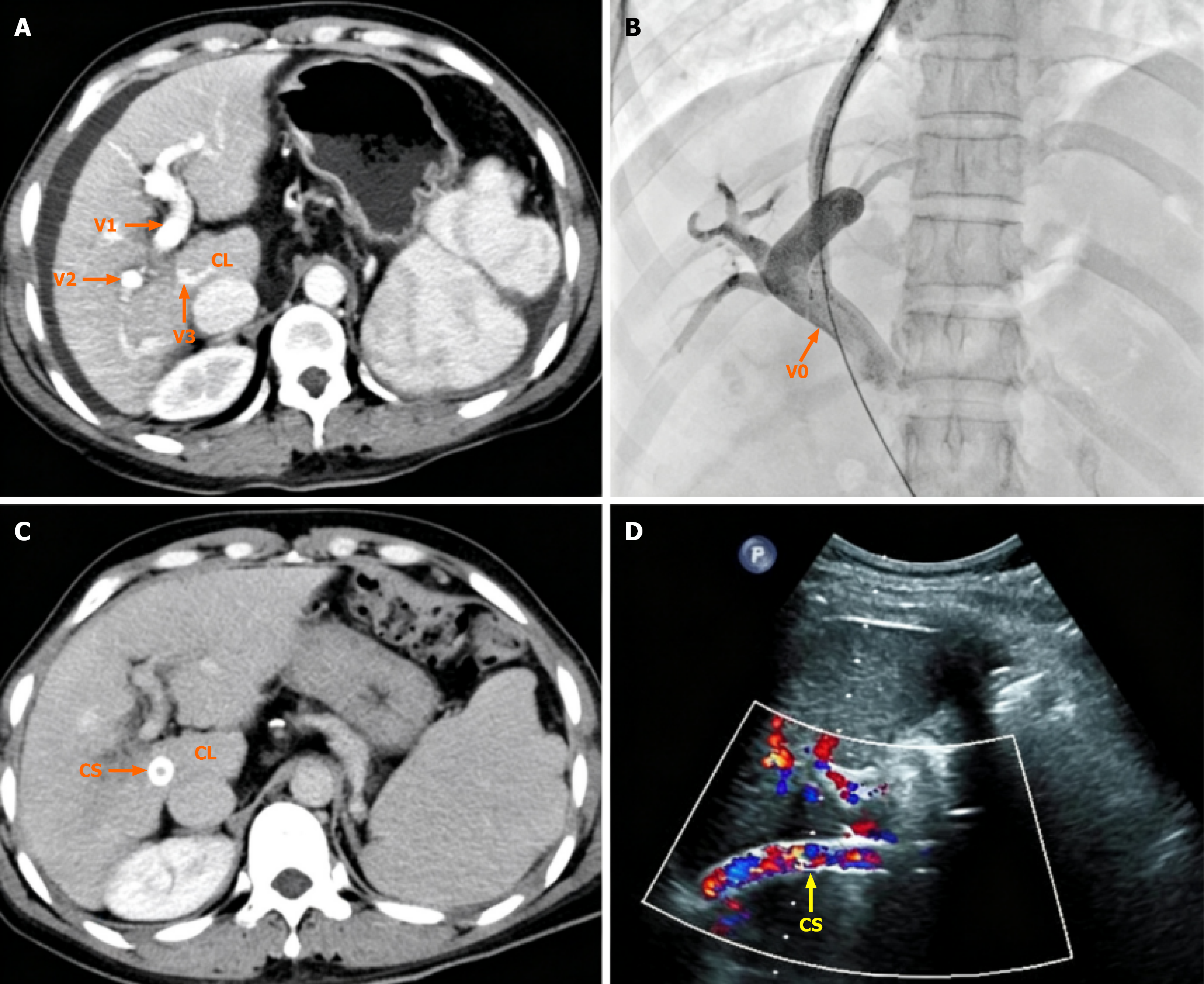
Figure 1 Case 1.
A: Operation procedures of transjugular intrahepatic portosystemic shunt; The preoperative computed tomography shows the thickened caudate lobe portal vein (CLPV); B: The stent enters the portal vein trunk via the CLPV; C: The stent was located in the CLPV, and the caudate lobe had no obvious atrophy; D: The color Doppler ultrasound examination revealed good blood flow filling in the stent within the CLPV. V0: Main portal vein trunk; V1: Left portal vein branch; V2: Right portal vein branch; V3: CLPV; CS: Covered stent; CL: Hepatic caudate lobe.
- Citation: Xu T, Lai XH, Ye QX, Liu LG, Xu ZJ. Transjugular intrahepatic portosystemic shunt via the caudate lobe portal vein due to anatomic variation: Two case reports. World J Gastrointest Surg 2026; 18(8): 120285
- URL: https://www.wjgnet.com/1948-9366/full/v18/i8/120285.htm
- DOI: https://dx.doi.org/10.4240/wjgs.120285