
















Copyright: ©Author(s) 2026.
World J Gastrointest Surg. Jul 27, 2026; 18(7): 120634
Published online Jul 27, 2026. doi: 10.4240/wjgs.v18.i7.120634
Published online Jul 27, 2026. doi: 10.4240/wjgs.v18.i7.120634
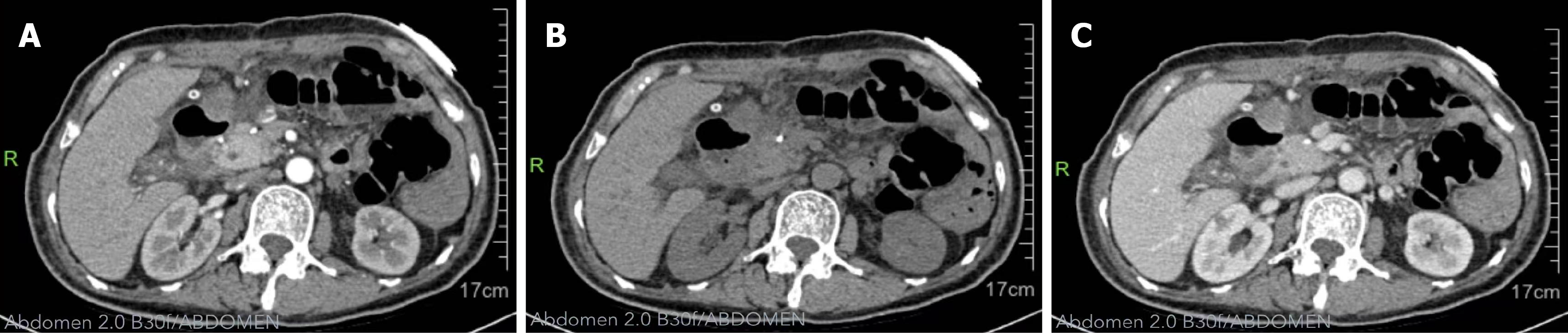
Figure 5 Postoperative contrast-enhanced abdominal computed tomography demonstrating changes following resection of the left-lobe liver metastasis, including postoperative exudation, localized effusion, gas accumulation, a turbid fat gap in the operative field, and a visible drainage tube in the upper abdomen.
A: Represents the arterial phase of the computed tomography (CT) scan; B: Non-contrast CT scan; C: Represents the venous phase of the CT scan.
- Citation: Yao JQ, Wu B, Yang JQ, Wang T, Shen YY. Liver metastasis from pancreatic neuroendocrine tumor: A case report. World J Gastrointest Surg 2026; 18(7): 120634
- URL: https://www.wjgnet.com/1948-9366/full/v18/i7/120634.htm
- DOI: https://dx.doi.org/10.4240/wjgs.v18.i7.120634