
















Copyright: ©Author(s) 2026.
World J Gastrointest Surg. Jul 27, 2026; 18(7): 120207
Published online Jul 27, 2026. doi: 10.4240/wjgs.v18.i7.120207
Published online Jul 27, 2026. doi: 10.4240/wjgs.v18.i7.120207
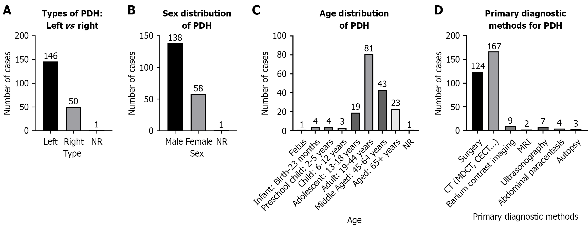
Figure 4 Epidemiological and clinical characteristics of paraduodenal hernia.
A: Paraduodenal hernia (PDH) showed a marked male predominance: 138 male cases vs 58 female cases, with 1 case of unclear gender, consistent with literature reports of male bias; B: Left-sided PDH was most common (n = 146), compared with 50 cases of right-sided PDH and 1 case of indeterminate laterality, reflecting anatomical predisposition to left-sided herniation; C: Highest incidence occurred in middle-aged adults (35-54 years, n = 81), followed by older adults (≥ 55 years, n = 43) and adolescents (13-18 years, n = 19). Cases were rare in infants and young children, suggesting a link to age-related abdominal changes or chronic intra-abdominal pressure; D: Cross-sectional imaging (computed tomo
- Citation: Chen LT, Pan BJ, Lu MD, Wang BH, Zhang N, Hu JW. Paraduodenal hernia with intestinal necrosis: Three case reports and review of literature. World J Gastrointest Surg 2026; 18(7): 120207
- URL: https://www.wjgnet.com/1948-9366/full/v18/i7/120207.htm
- DOI: https://dx.doi.org/10.4240/wjgs.v18.i7.120207