
















Copyright: ©Author(s) 2026.
World J Gastrointest Surg. Jul 27, 2026; 18(7): 120053
Published online Jul 27, 2026. doi: 10.4240/wjgs.120053
Published online Jul 27, 2026. doi: 10.4240/wjgs.120053
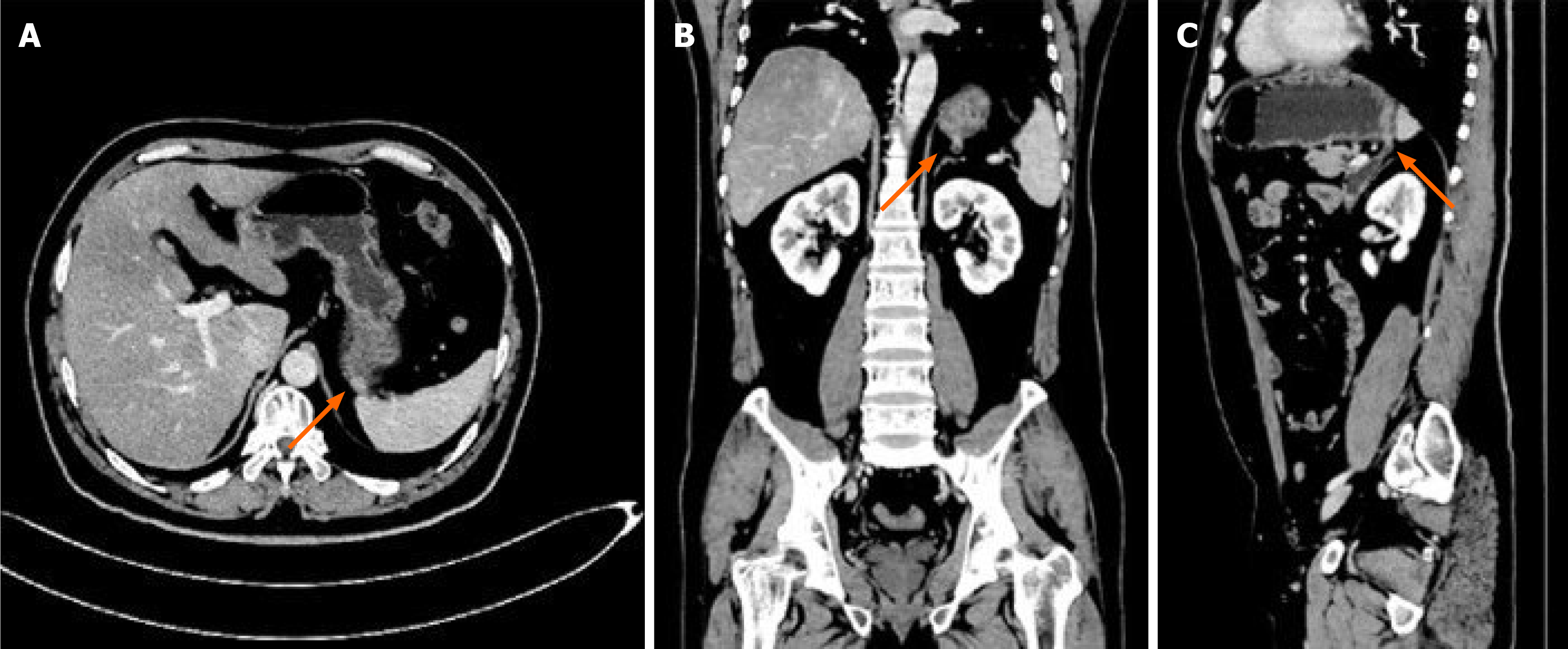
Figure 2 Contrast-enhanced computed tomography showing the fistula (arrows).
A: Axial view; B: Coronal view; C: Sagittal view. A focal defect in the posterior fundic wall communicates with an adjacent jejunal loop; fat planes are preserved, without inflammatory stranding.
- Citation: Chen XR, Guo HY, Liang R, Zhang CY, Yuan XB, Wang XF. Congenital gastrojejunal fistula in an asymptomatic adult: A case report. World J Gastrointest Surg 2026; 18(7): 120053
- URL: https://www.wjgnet.com/1948-9366/full/v18/i7/120053.htm
- DOI: https://dx.doi.org/10.4240/wjgs.120053