
















Copyright: ©Author(s) 2026.
World J Gastrointest Surg. Jul 27, 2026; 18(7): 119186
Published online Jul 27, 2026. doi: 10.4240/wjgs.v18.i7.119186
Published online Jul 27, 2026. doi: 10.4240/wjgs.v18.i7.119186
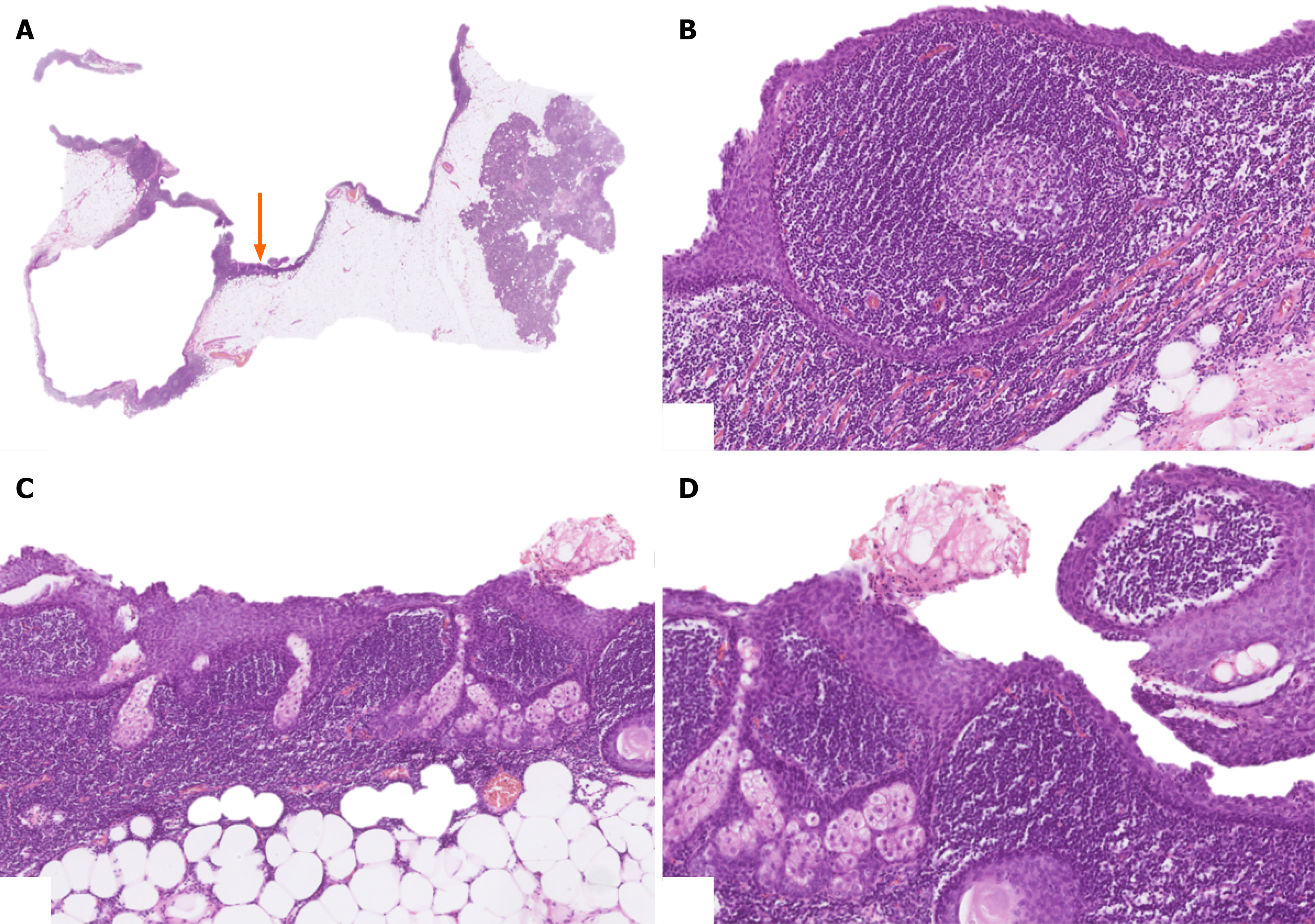
Figure 4 Lymphoepithelial cyst of the pancreas with sebaceous differentiation.
A: The cyst was localized in the pancreatic tail. The arrow depicts the area with sebaceous differentiation (5 × magnification); B: The cyst was lined by stratified squamous epithelium. Note the dense lymphatic tissue with formation of germinal centers in the cyst wall (80 × magnification); C: Numerous mature sebaceous lobules were present within the stratified squamous epithelium (40 × magnification); D: In addition to mature sebaceous glands, focal formation of sebaceous ducts was also present (80 × magnification).
- Citation: Badovinac D, Luzar B. Pancreatic lymphoepithelial cyst with sebaceous differentiation-encountering and management of these rare tumours: A case report and review of literature. World J Gastrointest Surg 2026; 18(7): 119186
- URL: https://www.wjgnet.com/1948-9366/full/v18/i7/119186.htm
- DOI: https://dx.doi.org/10.4240/wjgs.v18.i7.119186