
















Copyright: ©Author(s) 2026.
World J Gastrointest Surg. Jul 27, 2026; 18(7): 117966
Published online Jul 27, 2026. doi: 10.4240/wjgs.117966
Published online Jul 27, 2026. doi: 10.4240/wjgs.117966
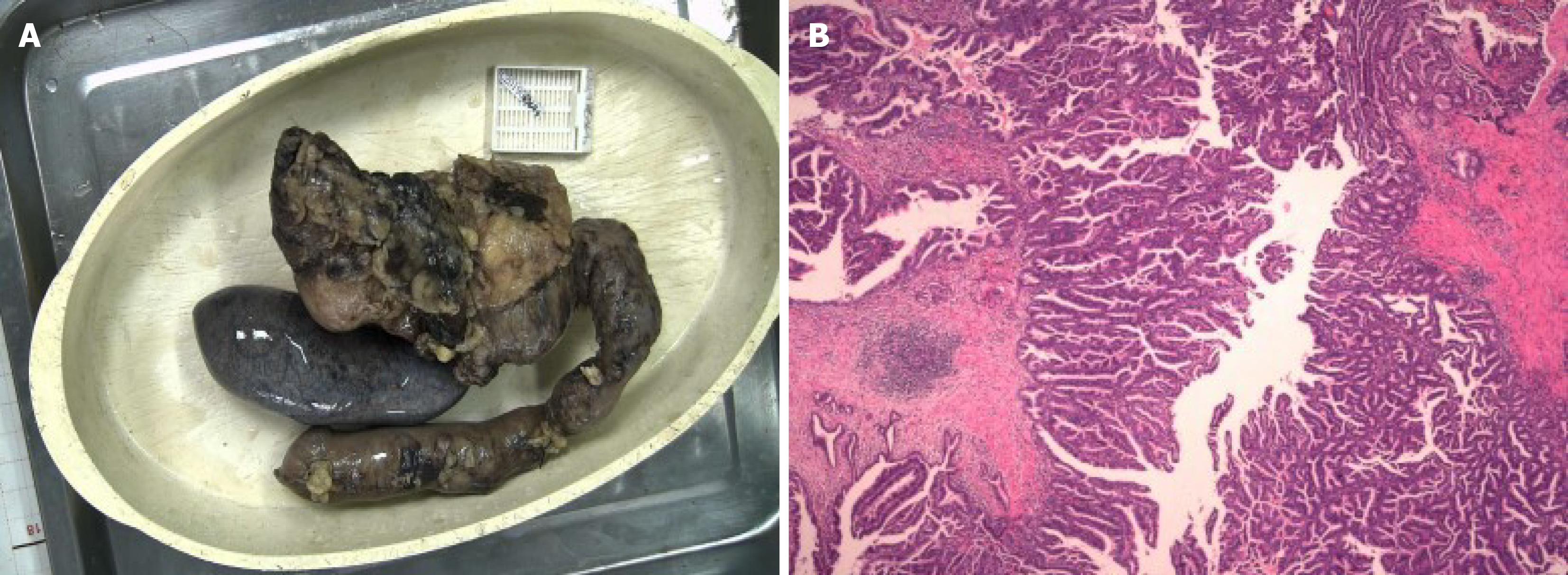
Figure 2 Pathological findings of duodenal papillary adenocarcinoma.
A: A gray-red solid mass (1.00 cm × 1.00 cm × 0.50 cm) at the duodenal papilla with a gray-white cut surface and moderate consistency, poorly demarcated from surrounding tissues. Attached organs include the stomach (8.00 cm × 6.50 cm × 4.00 cm), duodenum (length 27.00 cm), gallbladder (11.00 cm × 4.00 cm × 3.50 cm; rough mucosa, smooth serosa), and pancreas (7.50 cm × 6.00 cm × 2.00 cm; lobulated cut surface). No tumor invasion is observed in any organ; B: Papillary adenocarcinoma infiltrating the muscularis propria. No cancer infiltration is seen in the serosa or periserosal tissues.
- Citation: Li KN, Chen Q, Huang CT, Liu TQ, Li HF. Long-term survival after pancreaticoduodenectomy for duodenal papillary adenocarcinoma in a patient with acquired immunodeficiency syndrome: A case report and review of literature. World J Gastrointest Surg 2026; 18(7): 117966
- URL: https://www.wjgnet.com/1948-9366/full/v18/i7/117966.htm
- DOI: https://dx.doi.org/10.4240/wjgs.117966