
















Copyright: ©Author(s) 2026.
World J Gastrointest Surg. Jul 27, 2026; 18(7): 117966
Published online Jul 27, 2026. doi: 10.4240/wjgs.117966
Published online Jul 27, 2026. doi: 10.4240/wjgs.117966
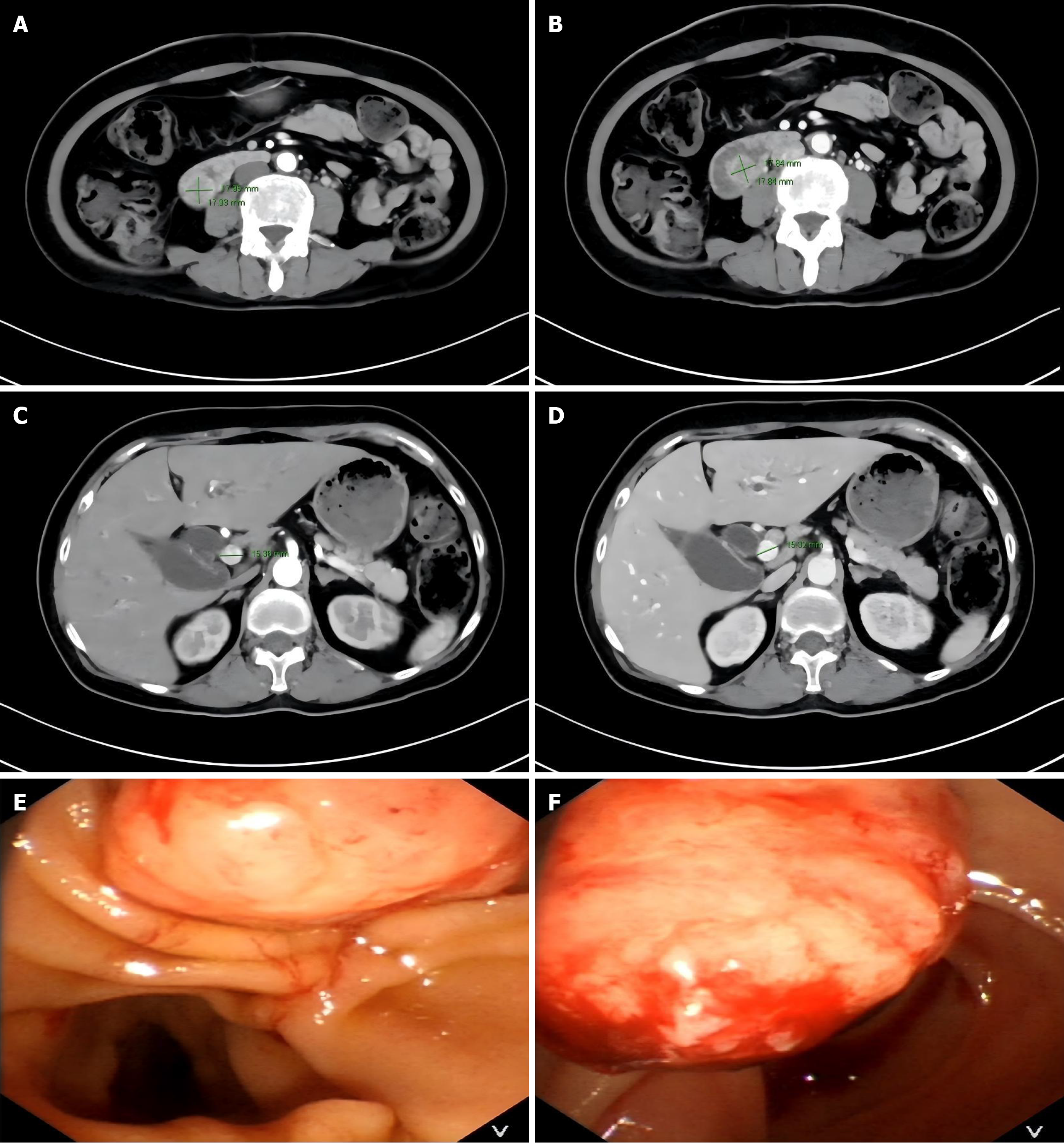
Figure 1 Imaging features of a duodenal papillary tumor.
A and B: Axial contrast-enhanced computed tomography shows an enhancing nodular lesion (1.80 cm × 1.80 cm) in the duodenal papilla region; C and D: Note the dilated biliary and pancreatic ducts (common hepatic duct: 1.50 cm) and the double duct sign; E and F: Gastroscopy reveals a tumor (1.50 cm × 1.50 cm) in the descending duodenum obscuring the nipple opening. The discrepancy between endoscopic (1.50 cm × 1.50 cm) and pathological (1.00 cm × 1.00 cm × 0.50 cm) tumor dimensions may be attributed to tissue shrinkage during formalin fixation, partial tissue loss from preoperative biopsy, and intraoperative manipulation.
- Citation: Li KN, Chen Q, Huang CT, Liu TQ, Li HF. Long-term survival after pancreaticoduodenectomy for duodenal papillary adenocarcinoma in a patient with acquired immunodeficiency syndrome: A case report and review of literature. World J Gastrointest Surg 2026; 18(7): 117966
- URL: https://www.wjgnet.com/1948-9366/full/v18/i7/117966.htm
- DOI: https://dx.doi.org/10.4240/wjgs.117966