
















Copyright: ©Author(s) 2026.
World J Gastrointest Surg. Jun 27, 2026; 18(6): 119246
Published online Jun 27, 2026. doi: 10.4240/wjgs.119246
Published online Jun 27, 2026. doi: 10.4240/wjgs.119246
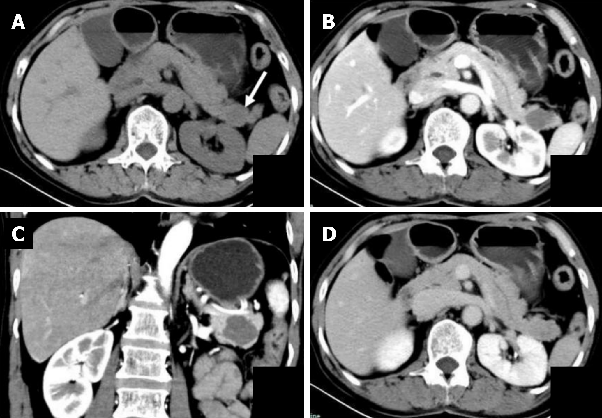
Figure 1 Preoperative computed tomography findings of the pancreatic schwannoma.
A: Plain computed tomography (CT) scan showing a well-marginated, slightly hypodense mass (arrow) in the pancreatic tail; B: Contrast-enhanced CT scans in the arterial phase demonstrating inhomogeneous mild enhancement of the lesion; C: Contrast-enhanced CT scans in the arterial phase demonstrating inhomogeneous mild enhancement of the lesion; D: Contrast-enhanced CT scan in the parenchymal phase showing progressive enhancement of the mass.
- Citation: Xu SM, Qiu LY. Incidental schwannoma of the pancreatic tail mimicking a solid pseudopapillary tumor: A case report. World J Gastrointest Surg 2026; 18(6): 119246
- URL: https://www.wjgnet.com/1948-9366/full/v18/i6/119246.htm
- DOI: https://dx.doi.org/10.4240/wjgs.119246