
















Copyright: ©Author(s) 2026.
World J Gastrointest Surg. Jun 27, 2026; 18(6): 119218
Published online Jun 27, 2026. doi: 10.4240/wjgs.119218
Published online Jun 27, 2026. doi: 10.4240/wjgs.119218
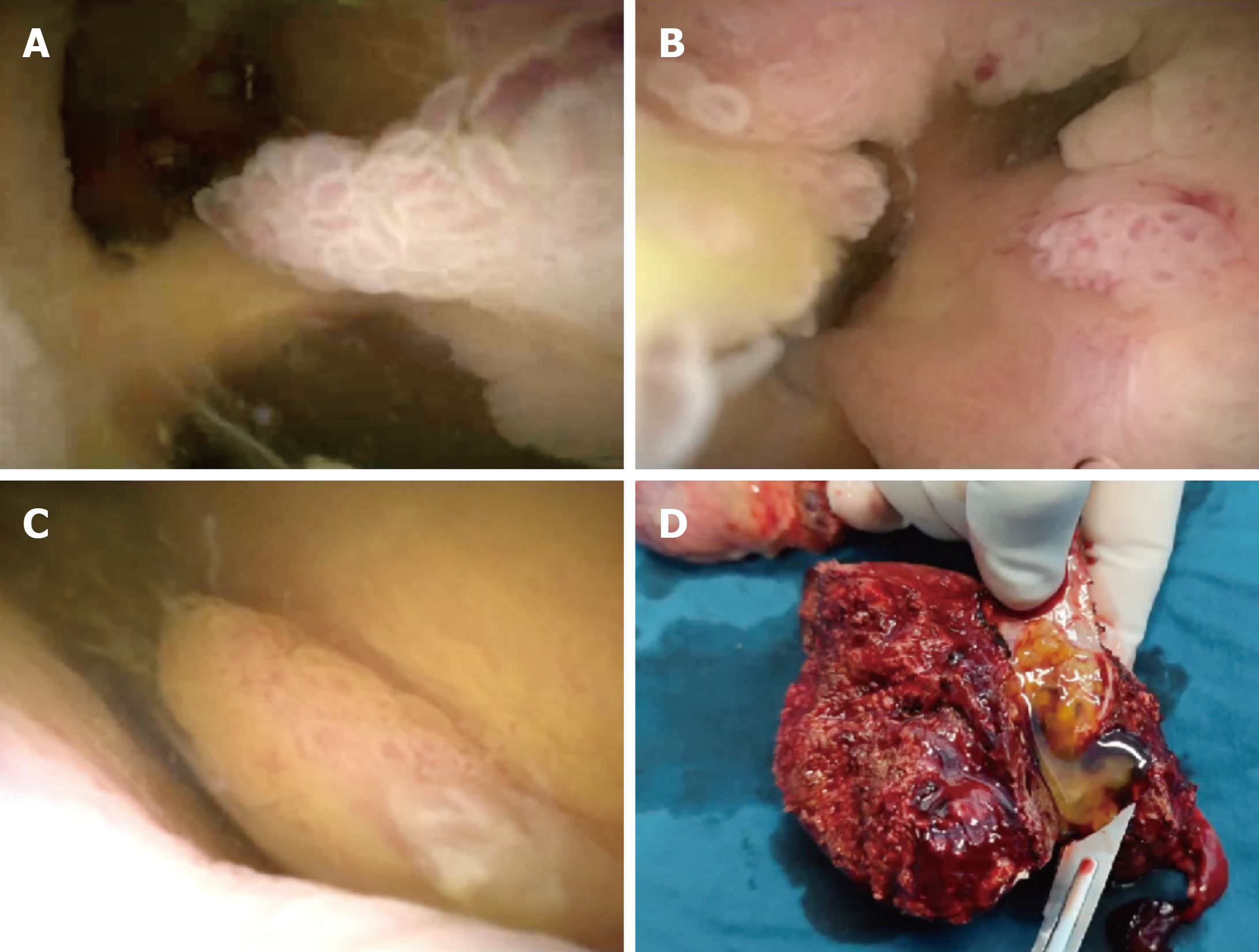
Figure 3 EyeMaxÔ direct visualization system.
A-C: Papillary eminence was found in the secondary branching bile duct of left outer lobe; D: Papillary neoplasm was located in the secondary bile duct of left outer lobe of liver (the gross specimen), and abundant gelatinous mucus was observed in the excised specimen.
- Citation: Fan WJ, Feng XX, Zhang ZC, Xiang S, Zhang WG, Wang Y, Liu M. Intraductal papillary neoplasm of the left intrahepatic bile duct found during early gastric cancer treatment: A case report. World J Gastrointest Surg 2026; 18(6): 119218
- URL: https://www.wjgnet.com/1948-9366/full/v18/i6/119218.htm
- DOI: https://dx.doi.org/10.4240/wjgs.119218