
















Copyright: ©Author(s) 2026.
World J Gastrointest Surg. Jun 27, 2026; 18(6): 119179
Published online Jun 27, 2026. doi: 10.4240/wjgs.119179
Published online Jun 27, 2026. doi: 10.4240/wjgs.119179
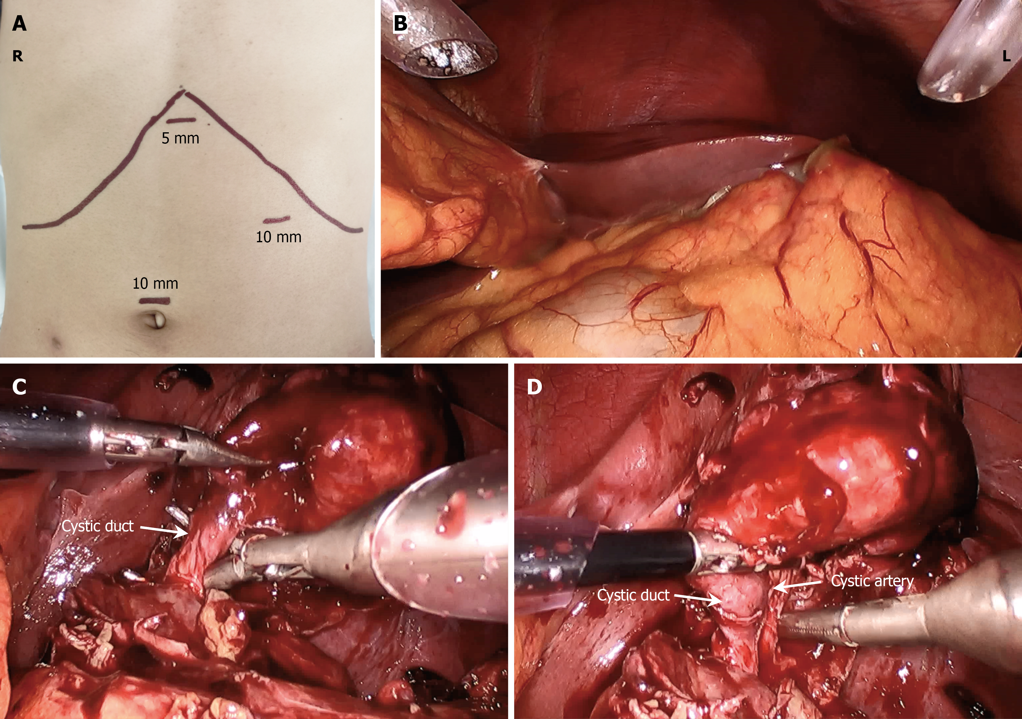
Figure 2 Illustrates the operative setup and key steps in the posterior dissection of Calot’s triangle.
A: Schematic diagram of laparoscopic port placement; B: Gallbladder located left of the falciform ligament with dense adhesions to the greater omentum; C: Posterior dissection of the cystic duct in Calot’s triangle; D: Posterior dissection of the cystic artery in Calot’s triangle.
- Citation: Chen ML, Ma X, Xiao L, Chen TT, Luo ZL, Xie XD. Posterior approach to Calot’s triangle in situs inversus totalis: A case report and review of literature. World J Gastrointest Surg 2026; 18(6): 119179
- URL: https://www.wjgnet.com/1948-9366/full/v18/i6/119179.htm
- DOI: https://dx.doi.org/10.4240/wjgs.119179